The anti apoptotic effect of leukotriene B4 in neutrophils
- 格式:pdf
- 大小:331.06 KB
- 文档页数:9
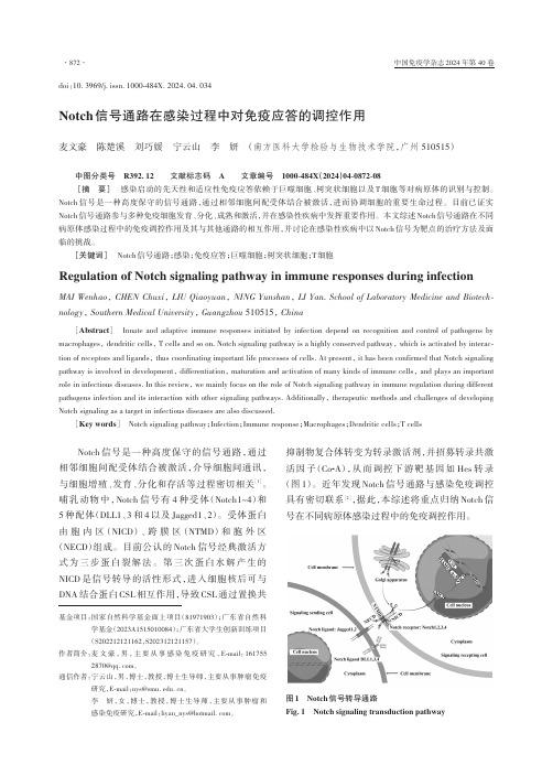
Notch信号通路在感染过程中对免疫应答的调控作用麦文豪陈楚溪刘巧媛宁云山李妍(南方医科大学检验与生物技术学院,广州 510515)中图分类号R392.12 文献标志码 A 文章编号1000-484X(2024)04-0872-08[摘要]感染启动的先天性和适应性免疫应答依赖于巨噬细胞、树突状细胞以及T细胞等对病原体的识别与控制。
Notch信号是一种高度保守的信号通路,通过相邻细胞间配受体结合被激活,进而协调细胞的重要生命过程。
目前已证实Notch信号通路参与多种免疫细胞发育、分化、成熟和激活,并在感染性疾病中发挥重要作用。
本文综述Notch信号通路在不同病原体感染过程中的免疫调控作用及其与其他通路的相互作用,并讨论在感染性疾病中以Notch信号为靶点的治疗方法及面临的挑战。
[关键词]Notch信号通路;感染;免疫应答;巨噬细胞;树突状细胞;T细胞Regulation of Notch signaling pathway in immune responses during infectionMAI Wenhao, CHEN Chuxi, LIU Qiaoyuan, NING Yunshan, LI Yan. School of Laboratory Medicine and Biotech⁃nology, Southern Medical University, Guangzhou 510515, China[Abstract]Innate and adaptive immune responses initiated by infection depend on recognition and control of pathogens by macrophages, dendritic cells, T cells and so on. Notch signaling pathway is a highly conserved pathway, which is activated by interac‑tion of receptors and ligands, thus coordinating important life processes of cells. At present, it has been confirmed that Notch signaling pathway is involved in development, differentiation, maturation and activation of many kinds of immune cells, and plays an important role in infectious diseases. In this review, we mainly focus on the role of Notch signaling pathway in immune regulation during different pathogens infection and its interaction with other signaling pathways. Additionally, therapeutic methods and challenges of developing Notch signaling as a target in infectious diseases are also discussed.[Key words]Notch signaling pathway;Infection;Immune response;Macrophages;Dendritic cells;T cellsNotch信号是一种高度保守的信号通路,通过相邻细胞间配受体结合被激活,介导细胞间通讯,与细胞增殖、发育、分化和存活等过程密切相关[1]。
雷公藤红素治疗类风湿关节炎的研究进展类风湿关节炎是一种慢性、自身免疫性疾病,主要影响关节和软组织,病变受多个因素的影响,而现有的治疗手段存在副作用和疗效较慢的问题。
近年来,雷公藤红素被发现在治疗类风湿关节炎方面具有良好的效果,以下是该领域的研究进展。
雷公藤红素(Triptolide)属于三萜类物质,是从雷公藤科植物雷公藤中提取出来的一种化合物。
研究表明,雷公藤红素具有多种药理作用,包括抗肿瘤、抗炎和免疫调节作用等。
在治疗类风湿关节炎方面,雷公藤红素的主要作用是通过抑制炎症反应和调节免疫系统来减轻病情。
研究表明,雷公藤红素可抑制多种炎症介质的产生,包括IL-1β、IL-6、IL-17和TNF-α等,从而减轻炎症反应,缓解关节疼痛和肿胀。
此外,雷公藤红素还能够影响B细胞和T细胞的功能,抑制其对自身抗原的反应,从而减少自身免疫反应,保护关节和软组织。
近年来的临床研究表明,雷公藤红素在治疗类风湿关节炎方面具有良好的效果。
一项关于雷公藤红素治疗类风湿关节炎的Meta分析表明,与常规治疗相比,雷公藤红素能够显著减轻关节疼痛和肿胀,并提高患者的生活质量。
尽管雷公藤红素的临床疗效已得到了广泛的认可,但其使用仍存在一定的风险和挑战。
雷公藤红素具有一定的毒性和副作用,如肝毒性、肾毒性和心血管毒性等。
因此,在使用雷公藤红素时,需要严格控制剂量,并结合其他药物进行联合治疗。
总之,雷公藤红素在治疗类风湿关节炎方面具有一定的临床疗效,并逐渐被广泛应用。
然而,当前对于雷公藤红素治疗类风湿关节炎的机制、剂量和疗程等方面仍存在许多未解决的问题,需要进一步的研究和探索。

三氧化二砷对Ⅱ型胶原诱导关节炎模型Treg和
Th17细胞亚群的影响的开题报告
研究背景:Ⅱ型胶原诱导关节炎(CIA)是一种常见的自身免疫疾病,其病理生理机制涉及到多种免疫细胞、细胞因子和信号通路。
最近的研
究表明,调节性T细胞(Treg)和Th17细胞亚群在CIA中发挥着至关重要的作用。
三氧化二砷(ATO)是一种常用的抗肿瘤药物,但其对免疫系统的影响尚未被充分研究。
研究目的:本研究旨在探讨ATO对CIA模型小鼠Treg和Th17细胞亚群的影响,以及其在CIA免疫病理中的作用机制。
研究方法:将大鼠分为对照组、CIA模型组和ATO处理组。
CIA模
型组和ATO处理组均注射Ⅱ型胶原免疫诱导关节炎。
ATO处理组在注射CIA后每天腹腔注射ATO,对照组和CIA模型组注射等量生理盐水。
采
用流式细胞术分析ATO对Treg和Th17细胞亚群的影响,并使用ELISA
法和Real-time PCR等技术评估ATO对白细胞介素(IL)-17、IL-10和Foxp3等基因和蛋白的表达水平的影响。
研究预期结果:预计ATO能够显著改善CIA模型小鼠的关节炎症状,同时增加Treg细胞数量并减少Th17细胞数量。
同时,ATO还将显著影
响白细胞介素(IL)-17、IL-10和Foxp3等基因和蛋白的表达水平,从
而发挥对CIA的治疗作用。
研究意义:该研究将为进一步了解ATO在自身免疫性疾病中的免疫调节作用、阐明T细胞亚群在CIA免疫病理中的作用以及研究新型CIA
治疗药物提供科学依据。

不仅仅是保健品,特殊结构的胶原蛋白或可成为抗癌靶点原创药明康德药明康德2022-08-01 07:30发表于美国说起胶原蛋白,或许人们最先想到的是超市货架上琳琅满目的胶原蛋白保健品,事实上I型胶原蛋白是人体内含量最为丰富的蛋白质之一,广泛存在于骨骼、肌腱和皮肤组织中。
关于胶原蛋白补充剂在改善皮肤和关节健康中的作用仍旧充满争议,现在,科学家发现比起作为功效不明的保健品,胶原蛋白可能有一个更大的用处:抗击癌症。
最近发表在Cancer Cell的一篇研究发现癌细胞通过产生特异性结构的胶原蛋白,保护其免于机体免疫反应的伤害,而靶向破坏这种特定结构的胶原蛋白可以减少癌细胞增殖、提高免疫治疗的效力。
胶原蛋白作为细胞外基质的一部分,一般情况下它由两条α1链和一条α2链组装形成三螺旋蛋白结构。
然而,在研究人类胰腺癌细胞系时,研究人员发现这些癌细胞仅表达了编码α1链的基因(COL1a1),而相比之下胶原蛋白的“生产大户”成纤维细胞则可以同时表达这两种基因。
进一步的分析发现,癌细胞通过表观遗传调控手段,使编码α2链的基因(COL1a2)发生超甲基化来实现基因沉默,以此产生由三个α1链组成的癌症特异性胶原蛋白“同源三聚体”结构。
为探明这一特殊的蛋白结构对于癌细胞生存和增殖的意义,研究人员构建了COL1a1特异性敲除的胰腺癌小鼠模型,这种小鼠体内癌细胞的COL1a1基因处于沉默状态,故而癌细胞无法产生具有α1同源三聚体结构的胶原蛋白。
这种癌症特异性α1同源三聚体结构的缺失显著减少了癌细胞增殖并引发了癌症微生物组的重编程。
这些变化削弱了肿瘤免疫抑制作用,并伴随着T细胞浸润增加以及癌细胞数量的减少。
不仅如此,COL1a1敲除小鼠对于抗PD-1疗法的应答明显增强,这些发现提示,由三个α1链组成的胶原蛋白同源三聚体结构对于维持癌细胞增殖和免疫抑制作用至关重要,靶向破坏这种“同源三聚体”结构胶原蛋白可以显著提升免疫疗法的抗癌效力。
聚集素与肿瘤的研究进展综述审校聚集素(Clusterin,CLU)亦称异二聚体硫酸糖蛋白(sulfated glycoprotein-2,SGP-2),是一种前列腺雄激素抑制物。
1983年由Blaschuk等首次从山羊睾丸网液(ram fete testis fluid)中分离出来,因其支持细胞聚集而得名,于1992年统一命名为聚集素(Clusterin)。
其后逐渐发现在聚集素广泛存在于人类与各种动物的许多器官与组织中,如睾丸、附睾、肾脏、肝脏、肺脏、子宫、卵巢、乳腺及前列腺等器官, 在睾丸、附睾、肝、肾等脏器表达水平较高。
也几乎存在于包括血浆、乳汁、尿液、脑脊液和精液等所有体液中[1]。
聚集素蛋白在进化中高度保守,在氨基酸水平上不同动物间具有很好的同源性。
最近发现,聚集素在细胞凋亡、脂类的运输、细胞周期调、DNA损伤、细胞黏附、组织重塑中起重要作用[2]。
这些作用提示聚集素可能在各种恶性肿瘤的发生发展过程中起重要作用。
目前对聚集素的认识源于一些肿瘤细胞对聚集素的研究。
一、聚集素的结构功能及其基因定位人类的聚集素基因是单拷贝基因,定位于8p12-p21,由9个外显子组成,含有1651个碱基,编码449个氨基酸组成的多肽链。
研究也证实了聚集素在体内可发挥具有相互矛盾的双重作用,这可能与CLU在细胞内通过选择性剪接形成糖基化和非糖基化两种蛋白质亚型有关,即分泌型的clusterin(sCLU)和核型clusterin(nCLU) ,其中sCLU是生理条件下聚集素的主要存在形式。
sCLU是一种细胞保护因子。
sCLU是一种相对分子量为76~80ku的糖基化蛋白[3],对大多数细胞来说是一种具有细胞保护作用的因子。
sCLU的表达由全长聚集素mRNA的第一个AUG密码子开始翻译 ,表达相对分子量为60ku的sCLU蛋白前体。
该蛋白可在前导肽引导下定位于内质网,经过加工,暴露出其αβ位点,并在由内质网向高尔基体转运过程中进行糖基化。
1周博达1 侯超1 综述 王月丹1北京大学医学部基础医学院 100083Email: wangyuedan@摘要: 血管内皮生长因子通过与其受体结合发挥生物学作用,促进内皮细胞有丝分裂及其增殖分化、迁移,导致血管新生,形成血管翳,在类风湿关节炎的病理过程中发挥关键的作用。
对抗血管内皮生长因子及其受体,从而阻止新生血管的形成为治愈类风湿关节炎提供了新的途径。
关键词:类风湿性关节炎;血管内皮生长因子;受体;类风湿关节炎(rheumatoid arthritis, RA)是一种严重的自身免疫功能障碍性疾病,在世界范围内的平均患病率约1%,而我国约有400 万RA患者。
其临床表现以反复发作的多发性小关节炎和不同程度侵蚀性改变及进行性强硬和畸形为主,并有骨和骨骼肌萎缩,是一种致残率较高的疾病。
RA的病理特征为关节滑膜慢性炎症、软骨吸收、骨质破坏和骨纤维化,其中一个重要的病理生理过程是新生血管翳的形成,而新生血管的生成是产生和维持RA血管翳的重要标志。
在RA发展过程中,滑膜组织中存在大量的血管,出现了血源性细胞的浸润、增殖和水肿,在血管内皮细胞里层也出现了对细胞因子等作用的靶点,同时内皮细胞对这些因子的反应可以维持和促进RA病理过程的发展,特别是新血管的生成[1]。
近年来,人们已经认识到血管生成在RA的侵蚀和破坏过程中发挥了重要作用。
中断血管生成在类风湿性关节炎的治疗中有重要的意义。
RA患者的关节滑膜中,存在一些生长因子、细胞因子和化学增活素从而影响血管的生成,许多生物活性物质还能通过上调血管生成刺激素的表达从而发挥间接作用。
其中,血管内皮生长因子及其受体在血管新生过程中起着重要的作用。
一、关于血管内皮生长因子及其受体血管内皮生长因子(vascular endothelial growth factor,VEGF),又称为血管通透性因子(vascular permeability factor, VPF),于1989年由Ferrara等发现,属于血小板源生长因子(platelet-derived growth factor, PDGF) 家族成员,是一种相对分子量在34000-46000的同源二聚体糖蛋白,由两个亚基通过二硫键结合,其热稳定性高,序列高度保守。
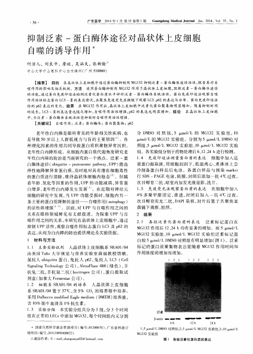

【高中生物】近代物理所揭示高LET射线诱导肿瘤细胞凋亡分子机理碳离子将恶性肿瘤细胞周期阻滞于g2/m期,抑制其生长,并明显诱导了肿瘤细胞凋亡。
中国科学院现代物理研究所放射医学系的研究人员利用兰州重离子研究所(HIRFL)提供的碳离子束,研究了高能线能量转移(let)射线诱导肿瘤细胞凋亡的分子机制,并获得了新发现。
细胞凋亡是电离辐射所致细胞死亡的主要形式。
p73是p53家族蛋白成员之一,在人类肿瘤细胞中很少发生缺失或突变,反而呈现出很高量的表达。
p73是抑制凋亡基因还是促进凋亡基因这个问题仍处于争论之中。
p73有两组蛋白异构体:tap73和np73。
tap73和δnp73被誉为肿瘤生死存亡的“开关”。
目前对于p73异构体在高let射线诱导的肿瘤细胞凋亡中的作用机制尚未见报道。
现代物理研究所放射医学系的研究人员发现,碳离子辐射诱导肿瘤细胞G2/M期阻滞,抑制其生长和增殖,并显著促进肿瘤细胞凋亡(如图1所示)。
其机制是电离辐射激活p73基因选择性剪接,启动p73介导的死亡受体和线粒体凋亡信号通路,进而促进肿瘤细胞凋亡的发生(如图2所示)。
此外,大蒜的天然活性产物二烯丙基二硫(DADS)不仅可以提高肿瘤细胞的放射敏感性,而且对正常细胞具有辐射防护作用。
进一步的实验证实,dads通过上调癌细胞TAp73/δNp73激活凋亡信号通路,促进癌细胞凋亡,与碳离子协同作用;对于正常细胞,TAp73下调/δNp73抑制其凋亡信号通路并促进DNA损伤的修复。
这些发现首次揭示了高LET辐射诱导肿瘤细胞凋亡的新分子机制,为提高重离子放射治疗的疗效和阐明其安全机制提供了新思路。
该研究得到国家自然科学基金委员会?中国科学院大科学装置联合基金重点项目和国家自然科学基金的资助。
研究结果发表在科学报告(,5:16020)和细胞周期(,DOI:10.1080/15384101..1104438)中。
芹菜素论文:芹菜素舌癌免疫组化凋亡【中文摘要】舌癌是最常见的口腔鳞状细胞癌,其临床表现为生长快,浸润性强,恶性程度高,常发生早期颈淋巴结转移。
目前,舌癌的治疗方案是以手术为主的综合序列治疗。
但是如果单纯手术治疗并不理想,尚存在局部容易复发、远处转移等难题。
因此,对于舌鳞状细胞癌,术后应采用副作用低的化疗药物进行联合治疗,将有助于降低复发率,提高生存率。
所以,对于恶性肿瘤术后化疗药物的研究就至关重要了。
芹菜素(Apigenin)是一种天然黄酮类化合物,存在于多种水果和蔬菜中,具有低毒、无诱变性及高效等特点。
国内外研究证实芹菜素可以抑制多种肿瘤的发生、发展,如消化道肿瘤、泌尿系统肿瘤以及生殖系统肿瘤等。
大量实验证明芹菜素在治疗恶性肿瘤方面的作用日益关注。
1.研究芹菜素对舌癌Tca8113细胞的生长抑制作用。
2.探讨芹菜素诱导肿瘤细胞凋亡、影响细胞增殖的机制。
3.研究芹菜素联合热疗方法对肿瘤细胞的影响,为芹菜素应用于治疗舌癌提供理论依据。
材料与方法:人工培养Tca8113细胞,配制不同质量浓度的芹菜素作用于该细胞;经过一定时间后,利用倒置显微镜观察Tca8113细胞的生长状态及形态学变化。
采用MTT法检测肿瘤细胞的芹菜素对Tca8113细胞的生长抑制作用;采用Annexin V-FITC/PI双染色法检测凋亡,而细胞周期进程的变化及凋亡率则采用流式细胞仪技术检测,统计学分析应用SPSS13.0软件,采用方差分析,卡方检验,卡方分割等方法检测。
取a=0.05为显著性检验水准。
结果:1.芹菜素对Tca8113细胞有明显的生长抑制作用:Tca8113细胞在各实验组浓度的芹菜素通过不同时间段作用后,其生长被抑制;且抑制作用的强度,随着芹菜素作用浓度的增高、作用时间的延长而增强。
2.倒置显微镜观察:40μmol/L芹菜素处理48h后,细胞体积变小、梭形散在、细胞境界清楚,贴壁能力减弱。
3.流式细胞仪检测结果表明,细胞在芹菜素的作用下,G1期比例逐渐增加,S期比例逐渐减少,细胞特异性停滞于G1/S期,与空白组比较差异有统计学意义(P<0.05)。
ADAMTSL5与银屑病发表时间:2018-04-19T13:10:31.387Z 来源:《医药前沿》2018年4月第12期作者:袁育林杨霞芳[导读] 可以成为银屑病中产生IL-17的CD8+ T细胞的活化抗原。
对ADAMTSL5的深入研究为阐明银屑病发病机制及靶向治疗带来了新希望。
(南宁市广西壮族自治区人民医院检验科广西南宁 530021)【中图分类号】R758.63 【文献标识码】A 【文章编号】2095-1752(2018)12-0014-03银屑病是一种常见的慢性复发性炎症性皮肤病。
其发病机制非常复杂,包括遗传、环境、免疫等多种因素参与其中。
虽然基于广泛的遗传,免疫和药理学证据,T细胞在银屑病发病机制中的作用已被广泛接受,但免疫系统在银屑病中被触发的机制仍然是一个迷。
银屑病易感基因座PSORS1上的HLA-C*06:02(6p21.33)是银屑病主要风险等位基因。
最近的研究显示ADAMTS样蛋白5(ADAMTSL5)作为Vα3S1/Vβ13S1TCR的HLA-C*06:02呈递的黑素细胞自身抗原,可导致产生IL-17的T细胞的活化,从而引起银屑病发病。
本文将对这一新鉴定的银屑病的自身抗原作简要综述。
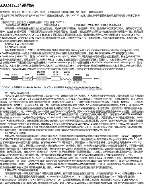
1.ADAMTSL5 的结构与功能1.1 ADAMTSL5结构含凝血酶敏感蛋白-1(TSP-1)基序的解聚蛋白样金属蛋白酶(a disintegrin-like and metalloproteinase with thrombospondin motifs,ADAMTS)超家族是一类整合于细胞外基质或游离于血浆中的基质金属蛋白酶亚家族,包括19种不同的ADAMTS蛋白[1]和至少7种ADAMTS-like(ADAMTSL)蛋白(ADAMTSL1-6和papilin)[2-3]。
ADAMTSL5是具有独特结构域的分泌型蛋白质,其包含N-末端TSR,富含半胱氨酸的模块,间隔基模块和C末端NTR模块,其通过富含脯氨酸的片段连接到间隔区(见图1)。
㊃综述㊃基金项目:2023年广州市科技局市校(院)联合资助项目肠道菌群代谢产物S C F A s 通过N F -κB 信号通路在帕金森病发病中的作用及机制研究(S L 2023A 03J 00590)通信作者:陈江瑛,E m a i l :1447369530@q q.c o m 胰高血糖素样肽-1在帕金森病肠-脑轴 中的作用研究进展周正新,梁秋雄,陈江瑛(暨南大学附属广州红十字会医院神经内科,广东广州510220) 摘 要:帕金森病(P D )是中老年时期常见的神经退行性疾病之一㊂P D 患者出现肠道炎症㊁便秘等胃肠道症状可能早于运动障碍㊂B r a a k 假说假设异常的α-突触核蛋白在肠道中开始积累,并通过迷走神经以类似朊病毒的方式传至大脑,且这一观点得到病理生理学证据的支持㊂P D 中 肠-脑轴 的关联在于代谢㊁免疫和炎症等相关机制,通过探讨其中机制可能成为治疗P D 的新思路㊂胰高血糖素样肽-1(G L P -1)作为研究较多的代谢物质之一,广泛参与在神经退行性病变中㊂本文对G L P -1在P D 肠-脑轴 中的作用进行综述,以期为P D 提供新的诊疗及研究思路㊂关键词:帕金森病;胰高血糖素样肽-1;肠-脑轴中图分类号:R 742.5 文献标志码:A 文章编号:1004-583X (2023)09-0855-04d o i :10.3969/j.i s s n .1004-583X.2023.09.015 帕金森病(P a r k i n s o n s d i s e a s e ,P D )是常见的运动障碍疾病,是一种表现有运动症状(运动迟缓㊁震颤㊁肌强直㊁步态异常)和非运动症状(胃肠功能紊乱㊁认知障碍㊁精神障碍㊁睡眠障碍和感觉障碍)的神经退行性疾病,其病因尚不完全清楚,亦缺乏根治手段㊂近年来, 肠-脑轴 这一概念的提出给包括P D 在内的神经疾病发病机制的探究提供了新思路㊂研究P D 与肠道系统相互联系的途径与其靶点,是丰富P D 研究的重点㊂2型糖尿病(t y pe 2d i a b e t e s m e l l i t u s ,T 2D M )是P D 发病的危险因素,且两者均与淀粉样多肽经过非正常折叠成纤维并随后聚集成淀粉样沉积有关[1]㊂因此,可以假设糖代谢异常可能是P D 的重要机制之一㊂P D 模型研究[2]表明,降糖药物胰高血糖素样肽-1(g l u c a g o n -l i k e p e p t i d e -1,G L P -1)受体激动剂具有神经保护作用㊂其中G L P -1,不只是一种可促进胰岛素信号传导㊁调节血糖水平且抑制食欲的肠促胰岛素激素[3],还是 肠-脑轴 的重要介质之一[4]㊂本文就G L P -1在P D肠-脑轴 中的作用进行综述,进一步探讨G L P -1与P D 的关系,以期为P D 的诊治提供新思路和依据㊂1 P D 的肠-脑轴 在大脑㊁肠道和肠道菌群存在着1种尚未被完全了解的交流系统,即 肠-脑轴 ㊂基于此轴,肠道菌群的改变被证明与一些神经疾病有关,如对P D 来说,肠道菌群的改变就是其突出的特征之一㊂1.1 病理联系 作为P D 黑质中形成的标志性聚集物,α-突触核蛋白(α-s y n u c l e i n ,α-s yn )不只在大脑中表达,且在肠道中也有表达[5];甚至其在肠道中的聚集比在大脑中病理形成更早[6]㊂B r a a k 等[7]提出,P D 是起源于中枢神经系统以外的病理模型㊂在该模型中,α-s yn 的错误折叠起自肠道,并通过迷走神经以朊病毒样的行为方式传至大脑[8]㊂且α-s yn 增加在P D 患者前驱阶段和早期无发病阶段的肠上皮中被发现[9]㊂可见,肠道中出现异常的α-s yn 发生在中枢神经退行性变的临床症状出现前㊂这表明,肠道可能是P D 发生的起源场所㊂虽然B r a a k 的观点备受争议,但中枢神经系统外α-s y n 错误折叠和聚集仍是P D 发病机制的经典假说,同时也是P D 肠-脑轴 的病理基础㊂1.2 肠道中介 细菌及其代谢产物除影响免疫系统外,还影响神经系统㊂F r i e d l a n d 等[10]提出了1个新的术语 m a p r a n o s i s 来描述微生物群相关的蛋白质病和神经炎症的过程( -o s i s )㊂尽管目前还未完全明确影响P D 的肠道菌群,但P D 的肠道状态和健康的肠道状态存在差异㊂临床研究[11]显示,P D 患者肠道出现异常改变,既有肠黏膜通透性增加㊁炎症迹象和受到细菌入侵,同时也有异常肠道菌群组成㊂在过表达α-s yn 小鼠大脑中小胶质细胞的激活和α-s yn 的病理形成及这些小鼠运动缺陷的产生都需肠道菌群参与,说明肠道菌群是P D 的危险因素[12]㊂P D 肠道菌群含有外膜脂多糖(l i p o p o l y s a c c h a r i d e s ,L P S )的细菌之相对丰度增加,而产生短链脂肪酸㊃558㊃‘临床荟萃“ 2023年9月20日第38卷第9期 C l i n i c a l F o c u s ,S e pt e m b e r 20,2023,V o l 38,N o .9(s h o r t-c h a i n f a t t y a c i d s,S C F A)的细菌之相对丰度降低,这是因为含有L P S的细菌与免疫激活和炎症有关,而产生S C F A的细菌与可能存在的抗炎性质有关[13]㊂一方面,L P S等促炎细菌成分激活小胶质细胞引发神经退行性变[14],暴露在L P S的全身效应随肠道屏障缺损(即肠道高渗透性)而增加,这是P D 的1个特征;另一方面,菌群的代谢产物S C F A与神经炎症的减轻㊁神经退行性变的减少和运动功能的改善有一定关系,在P D患者中这些菌群相对丰度往往减少[15]㊂位于肠道里的细菌代谢产物S C F A在P D的 肠-脑轴 假说中也发挥重要作用㊂S C F A的受体既存在于免疫细胞上,也存在于大脑细胞上,这为S C F A介导的 肠-脑轴 提供了物质联系[16]㊂目前,1个探索程度相对较低的机制就是S C F A对神经系统的影响㊂研究[17]发现,S C F A可引起G L P-1分泌,且对P D有神经保护作用㊂以上均提示,肠道菌群及其代谢产物对P D的产生有不可忽视的影响㊂2G L P-1在P D 肠-脑轴 中的作用肠-脑轴 与内分泌激素的产生与释放相关㊂研究[18]表明,T2D M是P D的危险因素,且胰岛素抵抗可能存在于大脑中,是神经退行性疾病的常见特征㊂作为与糖尿病相关的内分泌激素,G L P-1是肠促胰素信号系统成员之一,目前对其研究较为热门,其作用机制的研究最为深入也最为复杂㊂大量研究已证实,G L P-1广泛参与了P D的发生发展㊂可见,G L P-1可能在P D的 肠-脑轴 中发挥了重要作用㊂2.1 G L P-1受体在神经系统的分布 G L P-1作用于外周组织中的G L P-1受体(g l u c a g o n-l i k e p e p t i d e-1r e c e p t o r,G L P-1R)㊂这种受体遍布肠道㊁胃部㊁胰腺㊁心脏㊁肾脏㊁骨骼㊁血管和脂肪细胞[19]㊂G L P-1R 也广泛分布于中枢神经系统中,如与P D相关的黑质,其中也包括表达酪氨酸羟化酶(t y r o s i n e h y d r o x y l a s e,T H)的多巴胺能神经元[20],且神经元周围的小胶质细胞和星形胶质细胞也可表达G L P-1R[21],这些为G L P-1与神经退行性变的关系提供了物质基础㊂2.2肠道菌群对G L P-1的作用内源性G L P-1是由肠道L细胞产生㊂L细胞的数量沿胃肠道的纵轴逐渐增加;结肠腔内有大量肠道菌群,L细胞在其中比例最高,L细胞由于与肠道菌群直接接触而受到刺激㊂由此可见,肠道L细胞分泌的G L P-1受肠道菌群的影响;同时,其也受到细菌产生的S C F A影响㊂S C F A通过与L细胞上的跨膜游离脂肪酸受体(f r e e f a t t y a c i d r e c e p t o r2,F F A R2)结合发挥促分泌作用㊂L细胞中的法尼醇X受体(f a r n e s o i d Xr e c e p t o r, F X R)的激活通过减少F F A R2的表达和信号传导以减少S C F A诱导的G L P-1分泌;相反,S C F A诱导G L P-1分泌在敲除F X R小鼠中增强[17]㊂这提示,肠内分泌系统可能是调节G L P-1水平的主要调节位点,即对F X R的拮抗作用可能增强G L P-1的分泌㊂利用这种方法可增加G L P-1的产生,进而获得治疗益处㊂如前所述,P D肠道的特征是产生S C F A细菌丰度减低[15],这引起了各项研究对P D肠道G L P-1分泌的关注㊂因此,有研究[22]表明,与食用相同餐食的对照组相比,P D组餐后G L P-1水平较低㊂说明P D患者G L P-1分泌可能存在障碍,且这种障碍可能是由于产生S C F A的细菌较少所致㊂可以说,肠道菌群可通过G L P-1来影响大脑功能,即形成了 肠-脑轴 的联系㊂2.3 G L P-1对P D的作用 T2D M是P D的危险因素[1],G L P-1R激动剂能降低T2D M患者血糖[23]㊂这提示,G L P-1的稳态失调可能是P D的潜在机制㊂因此,存在将G L P-1信号的增强用于预防神经退行性病变的可能㊂研究[2]发现,G L P-1R激动剂恢复了合成多巴胺的限速酶T H在黑质的表达,还能进一步降低作为脂质过氧化产物和氧化应激标志物的4-羟基壬烯醛的产生[24],提高了神经营养因子,如神经胶质细胞系来源的神经营养因子和脑源性的神经营养因子的合成[25],并阻止体内T H阳性纹状体和中脑多巴胺能神经元中α-s y n[2,25]的积累㊂目前,已有研究进一步证明G L P-1R信号增强带来有益作用的机制㊂有研究[26]表明,G L P-1通过抑制核因子κB (n u c l e a r f a c t o rk a p p a-B,N F-κB)信号传导以发挥抗炎作用;而所对应的炎症正是在含有L P S的细菌的相对丰度增加和肠道屏障功能障碍下发生的㊂G L P-1R的激活通过调节细胞内线粒体活性氧(r e a c t i v e o x y g e n s p e c i e s,R O S)抑制P D的氧化应激(o x i d a t i v e s t r e s s,O S)㊂G L P-1R的激活增强了丝氨酸/苏氨酸激酶(t h r e o n i n e k i n a s e,A k t)的磷酸化并诱导转录因子环磷酸腺苷(c y c l i c a d e n o s i n e m o n o p h o s p h a t e,c AM P)反应元件结合蛋白[27],增强了抗凋亡B淋巴细胞瘤2(b-c e l l l y m p h o m a2,B c l-2)合成并降低促凋亡B c l-2相关X蛋白(b c l-2a s s o c i a t e d X p r o t e i n,B a x)[25,28]和细胞色素C (c y t o c h r o m eC,C y t C)[28]水平,使降低的B c l-2/B a x 比例正常化同时还降低了具有凋亡效应的天冬氨酸㊃658㊃‘临床荟萃“2023年9月20日第38卷第9期 C l i n i c a l F o c u s,S e p t e m b e r20,2023,V o l38,N o.9特异性半胱氨酸蛋白酶3(c a s p a s e3)水平[25]㊂这些研究为G L P-1对P D的作用提供了机制方面的支持证据㊂最近1项针对T2D M患者基于人群的纵向队列研究[29]结果显示,接受G L P-1R激动剂治疗组与其他降糖药物组比较,P D发病率降低㊂该研究为使用G L P-1R激动剂治疗的T2D M患者预防P D提供了可靠的流行病学证据,证明了G L P-1对P D神经保护和抗炎作用㊂3小结P D是发病率第2的神经退行性疾病,在全球范围内,P D导致的残疾和死亡人数高于其他神经系统疾病㊂目前的治疗方案仍未能满足临床需求,加之发病机制尚未完全明确,给P D患者预后带来极大挑战㊂作为目前研究最多且广泛参与T2D M和神经疾病发病机制中的肠促胰素,G L P-1为从肠道与神经系统角度分析 肠-脑轴 的形成开创了新的研究方向㊂期待未来会有更多关于G L P-1对P D 肠-脑轴 作用的研究,以更好地为防控P D等神经疾患奠定研究基础㊂参考文献:[1] N g u y e nP H,R a m a m o o r t h y A,S a h o o B R,e ta l.A m y l o i do l i g o m e r s:A j o i n t e x p e r i m e n t a l/c o m p u t a t i o n a l p e r s p e c t i v eo na l z h e i m e r'sd i s e a s e,p a r k i n s o n'sd i s e a s e,t y p e i i d i ab e t e s,a n da m y o t r o p h i c l a t e r a ls c l e r o s i s[J].C h e m R e v,2021,121(4):2545-2647.[2] Z h a n g L Y,J i nQ Q,Höl s c h e rC,e t a l.G l u c a g o n-l i k e p e p t i d e-1/g l u c o s e-d e p e n d e n ti n s u l i n o t r o p i c p o l y p e p t i d ed u a lr e c e p t o ra g o n i s tD A-C H5i s s u p e r i o r t o e x e n d i n-4i n p r o t e c t i n g n e u r o n si nt h e6-h y d r o x y d o p a m i n er a t p a r k i n s o n m o d e l[J].N e u r a lR e g e nR e s,2021,16(8):1660-1670.[3] K o p p K O,G l o t f e l t y E J,L iY,e ta l.G l u c a g o n-l i k e p e p t i d e-1(G L P-1)r e c e p t o r a g o n i s t s a n d n e u r o i n f l a mm a t i o n:i m p l i c a t i o n s f o r n e u r o d e g e n e r a t i v e d i s e a s e t r e a t m e n t[J].P h a r m a c o lR e s,2022,186:106550.[4] M a n f r e a d y R A,F o r s y t h C B,V o i g t R M,e ta l.G u t-b r a i nc o mm u n i c a t i o n i n p a r k i n s o n'sd i se a s e:E n t e r o e n d o c r i n er e g u l a t i o nb y G L P-1[J].C u r rN e u r o lN e u r o s c iR e p,2022,22(7):335-342.[5] L i u W,L i m K L,T a n E K.I n t e s t i n e-d e r i v e dα-s y n u c l e i ni n i t i a t e s a n da g g r a v a t e s p a t h o g e n e s i so f p a r k i n s o n'sd i s e a s e i nD r o s o p h i l a[J].T r a n s lN e u r o d e g e n e r,2022,11(1):44.[6] R o d r i g u e s P V,d e G o d o y J V P,B o s q u e B P,e t a l.T r a n s c e l l u l a r p r o p a g a t i o n o f f i b r i l l a rα-s y n u c l e i n f r o me n t e r o e n d o c r i n e t on e u r o n a lc e l l sr e q u i r e sc e l l-t o-c e l lc o n t a c ta n d i sR a b35-d e p e n d e n t[J].S c i R e p,2022,12(1):4168.[7] B r a a k H,Rüb U,G a i W P,e ta l.I d i o p a t h i c p a r k i n s o n'sd i se a s e:p o s s i b l er o u t e sb y w h i c hv u l n e r a b l en e u r o n a lt y p e sm a y b e s u b j e c t t o n e u r o i n v a s i o n b y a n u n k n o w n p a t h o g e n[J].JN e u r a lT r a n s m(V i e n n a),2003,110(5):517-536. [8] B o r g h a mm e r P.H o w d o e s p a r k i n s o n's d i s e a s e b e g i n?p e r s p e c t i v e s o n n e u r o a n a t o m i c a l p a t h w a y s,p r i o n s,a n dh i s t o l o g y[J].M o vD i s o r d,2018,33(1):48-57.[9]S h a n n o n KM,K e s h a v a r z i a n A,M u t l u E,e t a l.A l p h a-s y n u c l e i n i nc o l o n i cs u b m u c o s a i ne a r l y u n t r e a t e dP a r k i n s o n'sd i se a s e[J].M o v e m e n tD i s o r d e r s,2012,27(6):709-715.[10] F r i e d l a n dR P,C h a p m a nM R.T h e r o l e o fm i c r o b i a l a m y l o i d i nn e u r o d e g e n e r a t i o n[J].P L o S P a t h o g,2017,13(12): e1006654.[11] D u m i t r e s c uL,M a r t a D,D췍n췍u A,e ta l.S e r u m a n df e c a lm a r k e r s o f i n t e s t i n a l i n f l a mm a t i o n a n d i n t e s t i n a l b a r r i e r p e r m e a b i l i t y a r ee l e v a t e di n p a r k i n s o n's d i s e a s e[J].F r o n tN e u r o s c i.2021,18(15):689723.[12]S a m p s o n T,D e b e l i u sJ,T h r o n T,e ta l.G u t m i c r o b i o t ar e g u l a t em o t o rd e f i c i t sa n dn e u r o i n f l a mm a t i o ni na m o d e lo f p a r k i n s o n's d i s e a s e[J].C e l l,2016,167(6):1469-1480,e12.[13]J o e r s V,M a s i l a m o n i G,K e m p f D,e t a l.M i c r o g l i a,i n f l a mm a t i o na n d g u t m i c r o b i o t ar e s p o n s e si na p r o g r e s s i v em o n k e y m o d e lo f p a r k i n s o n's d i s e a s e:A c a s es e r i e s[J].N e u r o b i o lD i s,2020,144:105027.[14] A b d e l-H a q R,S c h l a c h e t z k i J C M,G l a s s C K,e t a l.M i c r o b i o m e-m i c r o g l i a c o n n e c t i o n sv i a t h e g u t-b r a i na x i s[J].JE x p M e d,2019,216(1):41-59.[15] N i s h i w a k iH,H a m a g u c h iT,I t o M,e ta l.S h o r t-c h a i nf a t t ya c i d-p r o d u c i n g g u t m i c r ob i o t ai s d ec r e a s ed i n p a r k i n s o n'sd i se a s eb u t n o t i nr a p i d-e y e-m o v e m e n t s l e e p b e h a v i o r d i s o r d e r[J].m S y s t e m s,2020,5(6):e00797-e007920.[16]S i l v aY P,B e r n a r d i A,F r o z z aR L.T h e r o l e o f s h o r t-c h a i n f a t t ya c i d sf r o m g u t m i c r ob i o t ai n g u t-b r a i nc o mm u n i c a t i o n[J].F r o n tE n d o c r i n o l(L a u s a n n e),2020,31(11):25.[17] D u c a s t e lS,T o u c h e V,T r a b e l s i M S,e t a l.T h e n u c l e a rr e c e p t o r F X R i n h i b i t s g l u c a g o n-l i k e p e p t i d e-1s e c r e t i o n i n r e s p o n s e t o m i c r o b i o t a-d e r i v e ds h o r t-c h a i nf a t t y a c i d s[J].S c iR e p,2020,10(1):174.[18] U y a r M,L e z i u s S,B u h m a n n C,e ta l.D i a b e t e s,g l y c a t e dh e m o g l o b i n(h b a1c),a n dn e u r o a x o n a l d a m a g e i n p a r k i n s o n'sd i se a s e(MA R K-P D s t u d y)[J].M o v D i s o r d,2022,37(6):1299-1304.[19] D r u c k e rD J.M e c h a n i s m s o f a c t i o na n d t h e r a p e u t i c a p p l i c a t i o no f g l u c a g o n-l i k e p e p t i d e-1[J].C e l lM e t a b,2018,27(4):740-756.[20] E l a b iO F,D a v i e sJ S,L a n eE L.L-d o p a-d e p e n d e n te f f e c t so fG L P-1R a g o n i s t s o n t h e s u r v i v a l o f d o p a m i n e r g i c c e l l st r a n s p l a n t e d i n t o a r a tm o d e l o f p a r k i n s o n d i s e a s e[J].I n t JM o l S c i,2021,22(22):12346.[21] C u iQ N,S t e i nL M,F o r t i nS M,e t a l.T h e r o l eo f g l i a i nt h ep h y s i o l o g y a n d p h a r m a c o l o g y o f g l u c a g o n-l i k e p e p t i d e-1:I m p l i c a t i o n s f o r o b e s i t y,d i a b e t e s,n e u r o d e g e n e r a t i o n a n dg l a u c o m a[J].B r JP h a r m a c o l,2022,179(4):715-726.㊃758㊃‘临床荟萃“2023年9月20日第38卷第9期 C l i n i c a l F o c u s,S e p t e m b e r20,2023,V o l38,N o.9[22] M a n f r e a d y R A,E n g e nP A,V e r h a g e n M L,e t a l.A t t e n u a t e dp o s t p r a n d i a lG L P-1r e s p o n s e i n p a r k i n s o n'sd i s e a s e[J].F r o n tN e u r o s c i,2021,2(15):660942.[23]郑鑫,朱育刚,王德峰.G L P-1受体激动剂对超重及肥胖2型糖尿病患者胰岛细胞功能影响的系统评价[J].临床荟萃, 2019,34(12):1102-1107.[24] Z h a n g Z Q,Höl s c h e r C.G I P h a sn e u r o p r o t e c t i v ee f f e c t si nA l z h e i m e r a n dP a r k i n s o n's d i s e a s em o d e l s[J].P e p t i d e s,2020,125:170184.[25] L vM,X u eG,C h e n g H,e t a l.T h eG L P-1/G I Pd u a l-r e c e p t o ra g o n i s tD A5-C Hi n h ib i t s t h eN F-κB i n f l a mm a t o r yp a t h w a y i nt h eM P T P m o u s em o d e l o f p a r k i n s o n's d i s e a s em o r e e f f e c t i v e l y t h a n t h e G L P-1s i n g l e-r e c e p t o r a g o n i s t N L Y01[J].B r a i nB e h a v,2021,11(8):e2231.[26] C h e nX,H u a n g Q,F e n g J,e ta l.G L P-1a l l e v i a t e s N L R P3i n f l a mm a s o m e-d e p e n d e n t i n f l a mm a t i o n i n p e r i v a s c u l a r a d i p o s et i s s u e b y i n h i b i t i n g t h e N F-κBs i g n a l l i n gp a t h w a y[J].JI n tM e dR e s,2021,49(2):300060521992981.[27]J a l e w a J,S h a r m aMK,G e n g l e r S,e t a l.An o v e lG L P-1/G I Pd u a l re c e p t o ra g o n i s t p r o t e c t sf r o m6-O H D Al e s i o ni nar a tm o d e l o f p a r k i n s o n's d i s e a s e[J].N e u r o p h a r m a c o l o g y,2017,1(117):238-248.[28] L iT,T u L,G u R,e ta l.N e u r o p r o t e c t i o no f G L P-1/G I Pr e c e p t o ra g o n i s t v i a i n h i b i t i o n㊁o f m i t o c h o n d r i a l s t r e s s b yA K T/J N K p a t h w a y i naP a r k i n s o n'sd i s e a s e m o d e l[J].L i f eS c i,2020,1(256):117824.[29] B r a u e rR,W e i L,M aT,e t a l.D i a b e t e sm e d i c a t i o n s a n d r i s ko f p a r k i n s o n's d i s e a s e:Ac o h o r t s t u d y o f p a t i e n t sw i t h d i a b e t e s[J].B r a i n,2020,143(10):3067-3076.收稿日期:2023-05-31编辑:张婷婷㊃858㊃‘临床荟萃“2023年9月20日第38卷第9期 C l i n i c a l F o c u s,S e p t e m b e r20,2023,V o l38,N o.9。

北京大学科技成果——海蛇降纤酶成果简介血栓形成是外科手术的常见并发症,也是现代介入性血管成形术后发生再阻塞的重要因素以及多种心脑血管疾病的致病、致死原因。
生理性血栓形成是止血的一种手段,而病理性血栓形成则可导致相关的脏器发生功能障碍。
目前心、脑部位的血栓性疾病己成为致残率与致死率最高的疾病之一,严重威胁到人类的健康。
据中国高血压联盟公布资料,我国高血压病人近1亿人,脑血管病患者450万人,每年新发生150万人,其中70%是缺血性脑血管病。
心肌梗死、心绞痛患者也有数百万人,还有大量的脑、心血管供血不足患者,四肢动、静脉血栓,眼底及内听动脉血管闭塞病人也需要抗凝和活血化瘀治疗。
蛇毒类凝血酶在体内具有较强的溶栓效果,它具有精氨酸脂酶活性,能够直接作用于纤维蛋白原,水解释放血纤肽2,导致纤维蛋白的单体首尾聚合而凝固,被称为类凝血酶。
但它在体内不激活凝血因子I,由它水解生成的纤维蛋白凝块不产生侧链交联,对纤溶酶的消化高度敏感,易被天然网状内皮系统或正常的纤溶作用所清除,因此导致胞浆中纤维蛋白原浓度显著下降,表现降纤、抗凝的效果。
临床上,蛇毒类凝血酶已成为防治血栓栓塞性疾病的有效药物。
纤维蛋白原是决定血液粘度的重要因素之一,类凝血酶减低了血浆中纤维蛋白原水平,从而降低了全血粘度及血浆粘度,增强了血流速度。
同时与凝血酶相反,类凝血酶不诱导血小板凝聚和释放,它们与富含血小板的血浆所形成的凝块不收缩,使机体能维持正常的止血功能。
国外已有Ancrod和Batroxobin蛇毒抗凝剂,国内则有五步蛇毒祛纤酶、东北白眉腹蛇抗栓酶(清栓酶)、江浙蝮蛇抗栓酶等用于临床。
虽然上述药物的名称不同但主要成分是一致的均为类凝血酶。
这些抗凝剂因具有祛纤、降粘、溶栓、解聚等独特的性质,已用于临床治疗脑血栓形成、脉管炎、治疗冠心病、心肌梗死,也有人用于治疗癌痛综合症。
目前认为抗栓酶是治疗脉管炎、深静脉炎、静脉栓塞形成最为理想的有效药物。
作者:李桢林江维王永红* 郝玉有储炬庄英萍张嗣良在合成培养基中分别添加缬氨酸(V al)、异亮氨酸(Ile)与亮氨酸(Leu)考察其对必特螺旋霉素生物合成的影响,证明V al、Ile及Leu对必特螺旋霉素多组分的合成有着不同的调节作用。
实验结果表明在36h分别添加0.5g/L的上述三种氨基酸,发酵液pH和铵离子浓度存在着显著差异,而V al脱氢酶活性都增加了2~3倍。
添加V al使菌体在60h之前糖耗速率加快,达到1.0g/(L?h),胞内丙酮酸积累,丙酮酸羧化酶活性增加20%~95%,柠檬酸合成酶活性降低41%,发酵液中丙酸和丁酸出现先增加后降低的现象,最终效价比对照高出45.3%,而总异戊酰组分则降低了23%,其他酰化组分降低了39.6%;添加Ile以后菌体糖耗速率降至对照的30%~40%,柠檬酸合成酶活性降低了47%,发酵液中的乙酸、丙酸、丁酸都出现大量积累的现象。
最终效价为对照的85%,而丙酰螺旋霉素III、乙酰螺旋霉素以及异丁酰螺旋霉素组分含量分别增加126%、50%、296%。
添加Leu对初级代谢影响不大,异戊酸在胞外积累,随后被重新吸收利用,最终异戊酰基螺旋霉素II和异戊酰基螺旋霉素III组分分别提高了41%和50%。
论文论文参考网同时总异戊酰基螺旋霉素相对含量也比对照提高了41.9%,效价和对照基本一致。
必特螺旋霉素;缬氨酸;亮氨酸;异亮氨酸;有机酸;铵离子ABSTRACT This paper demonstrated that supplementing valine (L-V al), isoleucine (L-Ile) and leucine (L-Leu) in synthetic media had different effects on biosynthesis of multi-components of biotechspiramycin. The results showed that the supplement of 0.05g/100ml amino acids after 36h of fermentation resulted in different pH values and ammonium ion concentrations of fermentation broth, and 2 to 3 time-increase of the activity of valine dehydrogenase. Within 60h after the supplement of L-V al, the glucose consumption rate was increased to 0.1 g/(100ml?h), pyruvic acid was accumulated inside the cells, the activity of pyruvate carboxylase (PC) was increased in the range of 20%~95%, the activity of citrate synthase (CS) was decreased by 41%, the propionic acid and butyric acid in the fermentation broth were increased at first and then decreased with the ultimate biological titer of biotechspiramycin being 45.3% higher than that of the control group, the relative content of total isovalerylspiramycin components were decreased by 23%, and the relative content of other acylated components was declined by 39.6%. After the supplement of L-Ile, the glucose consumption rate was decreased to 30%~40% then that of the control group, the activity of CS was decreased by 47%, large amounts of acetic acid, propionic acid and butyric acid in the fermentation b省钱屋购物省钱交流必备的网站在这里达人们会教你如何用80块钱充值100元钱话费的省钱计划。
曲古抑菌素A对胃癌BGC823细胞增殖及凋亡的影响姚洁【摘要】目的:研究曲古抑菌素A(TSA)对体外培养的胃癌BGC823细胞增殖及凋亡的影响.方法:体外培养人胃癌BGC823细胞,采用CCK8法测定不同浓度(100 nmol/L,200 nmol/L,300 nmol/L,400 nmol/L)及不同时间点(12 h、24 h、48 h)TSA对细胞增殖的影响.用流式细胞术检测48 h不同TSA浓度BGC823细胞的凋亡率.结果:CCK8法检测结果发现TSA对胃癌BGC823细胞有明显的增殖抑制作用,且呈浓度依赖性和时间依赖性.流式细胞术检测结果发现TSA可诱导人胃癌BGC823细胞凋亡,且呈浓度依赖性.结论:TSA可抑制体外培养的胃癌BGC823细胞生长,并可诱导其发生凋亡.【期刊名称】《临床医药实践》【年(卷),期】2019(028)003【总页数】3页(P182-183,240)【关键词】曲古抑菌素A;肿瘤;胃癌;增殖;凋亡【作者】姚洁【作者单位】山西医科大学附属太原中心医院,山西太原 030009【正文语种】中文【中图分类】R574.62胃癌到了进展期才能诊断,预后差[1]。
其治疗主要是外科手术,再辅助放、化疗以及药物治疗。
目前,治疗胃癌的药物大多基于体外杀死癌细胞的实验。
研究发现,组蛋白去乙酰化酶抑制剂可通过一系列非组蛋白乙酰化,转录激活肿瘤抑制基因和调节血管生成基因,从而对抗肿瘤发展[2]。
曲古抑菌素A(TSA)作为一种组蛋白去乙酰化酶及乙酰化酶抑制剂,为研究人员熟知。
目前研究认为TSA对肝癌[3]、肺癌[4]、胶质母细胞瘤[5]等均有抑制生长作用,但在胃癌中的研究较少见。
本文拟研究TSA对胃癌BGC823细胞的增殖及凋亡的影响,以期为TSA应用提供数据支持。
1 材料与方法1.1 材料胃癌BGC823细胞购自中国科学院细胞库。
TSA购自Sigma公司。
CCK8和RPMI-1640培养液购自武汉博士德生物工程有限公司。

替米沙坦对小鼠抗衰老klotho蛋白表达的影响及机制于凤芹;彭海;邢宏义;黎钢;李敏【期刊名称】《脑与神经疾病杂志》【年(卷),期】2011(019)001【摘要】Objective To investigate the effect and the mechanisms of Telmisartan on the expression of klotbo protein in the brain choroid plexus in mice. Methods 32 male Kunming mice were divided randomly into four groups: the nonnal control group, NOS blockade ( L-NAME) group. Telmisartan group and L-NAME plus Telmisartan group. All animals were administered by corresponding medicine for four weeks, respectively. The expression of klotho protein in the brain choroid plexus were measured by immunohistochemistry and Western blot technigues. The change of expression of RhoA/ROCK mRNA were detected by RT-PCR. Results Compared with the control group, the expression of RhoA and Rock mRNA increased significantly ( P<0. 01) and the expression of klotho protein decreased sharply ( P<0. 01) in the L-NAME group. Telmisartan reversed these changes( P<0. 01) .Conclusion These findings suggest that Telmisartan could intervene down-regulate the expressionof klotho protein induced by L-NAME. The mechanism might be related to inhibit the activities of Rho / ROCK signaling pathway.%目的观测替米沙坦对小鼠脑脉络丛klotho蛋白表达的影响及机制.方法 32只昆明小鼠,随机分为四组,即正常对照组、一氧化氮合酶(NOS)抑制组(L-NAME组),替米沙坦组和L-NAME+替米沙坦组,连续处理4周.免疫组织化学方法观察klotho蛋白表达;Western blot检测klotho蛋白表达水平的变化;RT-PCR检测RhoA和ROCK mRNA表达水平.结果与对照组比较,L-NAME组RhoA和ROCK mRNA表达升高(P<0.01);klotho蛋白表达明显降低(P< 0.01).给予替米沙坦后,RhoA和ROCK mRNA表达降低(P<0.01);klotho蛋白表达明显增高(P< 0.01).结论替米沙坦可干预L-NAME所致的klotho蛋白表达下调,其机制可能与抑制Rho/ ROCK信号通路活性有关.【总页数】4页(P42-45)【作者】于凤芹;彭海;邢宏义;黎钢;李敏【作者单位】430022,武汉,华中科技大学同济医学院附属协和医院神经内科;430022,武汉,华中科技大学同济医学院附属协和医院神经内科;430022,武汉,华中科技大学同济医学院附属协和医院神经内科;430022,武汉,华中科技大学同济医学院附属协和医院神经内科;湖北省宜昌市第二人民医院神经内科【正文语种】中文【中图分类】R741.02【相关文献】1.银杏叶提取物对衰老小鼠脑组织Klotho蛋白表达的影响及机制 [J], 邓刚;彭海;杨艳飞;于凤芹2.束缚应激对小鼠血浆α-klotho蛋白及肾脏α-klotho mRNA表达的影响 [J], 杨伟;苏显明;王颖;王瑛;马奕3.益髓灸对D-半乳糖衰老小鼠模型海马组织Klotho蛋白表达及胰岛素通路的影响[J], 赵利华;陈望龙;吴萍萍;陈树燕;罗志洪;方玉丽4.\"Becn1-BCL2-Klotho轴\"——自噬抗衰老新机制 [J], 王炳蔚;唐致恒;郑瑞茂5.抗衰老Klotho蛋白对过氧化氢诱导的人脐静脉内皮细胞氧化损伤的保护作用及其机制 [J], 王均鹏;孟丽霞;潘黎明;吕云波;李俊明因版权原因,仅展示原文概要,查看原文内容请购买。

蛋白酶抑制剂leupeptin增强抗肿瘤药物对肿瘤细胞的增殖抑制作用王莹;吴淑英;甄永苏【期刊名称】《医学研究杂志》【年(卷),期】2012(41)10【摘要】Objective To observe the modulating effect of leupeptin, a protease inhibitor, on the anti - proliferation efficacy of anti-cancer drugs including taxol (TAX) , gemcitabine ( GEM) , and pingyangmycin (PYM) in cancer cells. Methods Mammary carcinoma MCF -7 cells and pulmonary carcinoma PG cells were exposed to various concentrations of leupeptin alone or in combination with taxol ( TAX) , gemcitabine ( EM) , and pingyangmycin ( PYM ) , respectively. After 48 hours exposure, cell proliferation was determined by MTT assay. Flow cytometry was used to analyze cell cycle distribution. Results At the concentration of 50祄oI/L or higher, leupeptin enhanced the inhibitory effect of those three anticancer drugs on the proliferation of both cell line. More marked enhancement was found in the combination of leupeptin and TAX. MCF - 7 cell cycle distributions were changed at the low dosage GEM or TAX combination with leupeptin. Conclusion Leupeptin could enhance the inhibition effect of anticancer drugs, in particular, taxol, on cancer cells. The efficacy may be varied in different cell lines and drugs.%目的研究蛋白酶抑制剂(protease inhibitor) leupeptin对化疗药物紫杉醇(taxol,TAX)、吉西他滨(gemcitabine,GEM)以及平阳霉素(pingyangmycin,PYM)在体外抑制肿瘤细胞增殖作用的影响.方法选取一定浓度梯度的酶抑制剂leupeptin单独或联合TAX、GEM和PYM与肿瘤细胞孵育,48h后采用MTT法测定肿瘤细胞生长的变化并与相应浓度的TAX、GEM和PYM单药组相比较.采用流式细胞术测定MCF-7细胞周期分布.结果 leupeptin在浓度高于50μmol/L时可增强化疗药物对肺癌PG细胞和乳腺癌MCF-7细胞增殖的抑制作用.leupeptin对TAX的增效作用尤为显著.leupeptin与低浓度的GEM和TAX联合应用可改变MCF-7的细胞周期分布.结论 leupeptin可增强化疗药物对肿瘤细胞增殖的抑制作用,但其作用程度与肿瘤细胞株的种类、化疗药物的种类及leupeptin的浓度均相关.【总页数】5页(P56-60)【作者】王莹;吴淑英;甄永苏【作者单位】100050 中国医学科学院/北京协和医学院医药生物技术研究所;100050 中国医学科学院/北京协和医学院医药生物技术研究所;100050 中国医学科学院/北京协和医学院医药生物技术研究所【正文语种】中文【相关文献】1.干扰Nrf2可增强HirsutanolsA对肿瘤细胞增殖的抑制作用 [J], 马建国;李厚金;邓蓉;冯公侃;朱孝峰2.苦荞胰蛋白酶抑制剂对HL-60细胞增殖的抑制作用 [J], 王宏伟;乔振华;任文英;鹿育晋;朱镭;张丽;张政;王转花3.木犀草素抗肿瘤细胞增殖及增敏抗肿瘤药物作用研究 [J], 王洪燕;全康;蒋燕灵;吴加国;唐修文4.抗肿瘤药物联合磁场在抑制肿瘤细胞增殖中的应用研究 [J], 徐玲玲;黄林洁;洪文婷;赵铁军;方金勇5.胰蛋白酶抑制剂联合抗肿瘤药物对小鼠Lewis肺癌转移的抑制作用 [J], 李克生;杜惠芬;施苓;郭红云;唐清秀;王兰因版权原因,仅展示原文概要,查看原文内容请购买。

标题:雷公藤甲素诱导的caspase(含半胱氨酸的天冬氨酸蛋白水解酶)依赖的细胞死亡通过介导在白血病细胞线粒体途径引言:雷公藤甲素诱导的caspase依赖的细胞凋亡通过线粒体介导的通路在白血病细胞雷公藤甲素,从分离出一种二萜中国herbTripterygium wilfordiiHook.f,表现出抗肿瘤活广泛一系列实体瘤。
在这里,我们研究它的对白血病细胞和效果发现,在100纳米或更小,它有效诱导凋亡的各种白血病细胞系和原发性急性髓性白血病(AML)的原始细胞。
然后,我们试图找出其作用机制。
伴随着雷公藤甲素诱导caspasedependent细胞死亡显著降低XIAP水平。
ForcedXIAP表达减弱triptolideinduced细胞死亡。
雷公藤甲素也减少Mcl-1的,但不BCL-2和Bcl-XLlevels。
Bcl-2的过度抑制triptolideinduced凋亡。
线粒体膜此外,雷公藤甲素引起的损耗潜力和细胞色素C释放。
caspase-9的基因敲除细胞耐药,同时的caspase-8缺失细胞对雷公藤甲素敏感,暗示的关键性线粒体但没有死亡受体途径雷公藤甲素诱导的细胞凋亡。
雷公藤甲素也增强了细胞死亡诱导其他抗癌剂。
总的来说,我们的结果表明,雷公藤减小XIAP和强效诱导caspasedependent凋亡通过线粒体途径介导在白血病细胞低纳摩尔浓度。
强效雷公藤甲素的体外抗白血病活性值得进一步研究这种化合物对白血病的治疗和其他恶性肿瘤。
介绍:雷公藤Hook.f中,卫矛科的一员植物家族,已用于中国药世纪。
雷公藤内酯醇,二萜类化合物,首次从植物和结构上的特点在1972年并已被用于治疗各种自身免疫疾病的并作为患者的器官和组织移植的免疫抑制剂。
近日,雷公藤甲素所表现出具有抗肿瘤特性范围广泛的抑制生长和诱导细胞凋亡人肿瘤细胞。
雷公藤也显示敏感细胞死亡诱导的各种试剂,如Apo2/Trail ,TNF-α,和不同的化疗药物。
The anti-apoptotic effect of leukotriene B 4in neutrophils:A role for phosphatidylinositol 3-kinase,extracellularsignal-regulated kinase and Mcl-1Darlaine Pe ´trin,Sylvie Turcotte,Annie-Kim Gilbert,Marek Rola-Pleszczynski,Jana Stankova *Immunology Division,Department of Pediatrics,Faculty of Medicine,Universite ´de Sherbrooke,3001,North 12th Avenue,Sherbrooke,Que ´bec,Canada J1H 5N4Received 25April 2005;accepted 24May 2005Available online 20June 2005AbstractThe constitutive commitment of neutrophils to apoptosis is a key process for the control and resolution of inflammation and it can be delayed by various inflammatory mediators including leukotriene B 4(LTB 4).The mechanisms by which LTB 4contributes to neutrophil survival are still unclear and the present work aims at identifying intracellular pathways underlying this effect.Inhibition of human neutrophil apoptosis by LTB 4was abrogated by the phosphatidylinositol 3-kinase (PI3-K)inhibitor wortmannin and by the specific MEK inhibitor PD98059.In contrast,inhibitors of p38MAPK,Jak2/3and Src did not hinder the anti-apoptotic effect of LTB 4.We also investigated the effects of members of the Bcl-2family as they play a crucial role in the regulation of programmed cell death.When neutrophils were incubated with LTB 4for 1to 6h,the mRNA levels of the anti-apoptotic protein Mcl-1were upregulated approximately 2-fold,while those of the pro-apoptotic protein Bax were downregulated 3-to 4-fold,as determined by real-time PCR.Accordingly,Western blot analysis revealed that the expression of Mcl-1was upregulated in presence of LTB 4,while flow cytometric analysis revealed that Bax protein was downregulated.Furthermore,the modulatory effects of LTB 4on Mcl-1and Bax proteins were abolished in the presence of either wortmannin or PD98059.Taken together,these results demonstrate the participation of PI3-K and MEK/ERK kinases,as well as regulatory apoptotic proteins such as Mcl-1and Bax,in the anti-apoptotic effects of LTB 4in human neutrophils.D 2005Elsevier Inc.All rights reserved.Keywords:Human neutrophils;Lipid mediators;Apoptosis;Signal transduction;Leukotriens;Mcl-11.IntroductionNeutrophils,also known as polymorphonuclear leuko-cytes (PMN)3,represent a first line of defense againstinvading microorganisms.These phagocytic cells are key players of inflammation,rapidly responding to chemotactic agents and producing additional inflammatory mediators [1,2].Neutrophils have a very short half-life (8–20h)as they undergo constitutive apoptosis,a process assuring the elimination of dead cells without the release of cytotoxic granules into the surrounding tissues.Among the different inflammatory mediators,leuko-triene B 4(LTB 4)is well recognized for its ability to delay neutrophil apoptosis [3–5].However,the mechanisms underlying this anti-apoptotic effect are not clear.LTB 4is a lipid mediator derived from nuclear membrane phospholipids via the action of 5-lipoxygenase [6].LTB 4is rapidly synthesized by neutrophils and alveolar macro-0898-6568/$-see front matter D 2005Elsevier Inc.All rights reserved.doi:10.1016/j.cellsig.2005.05.021Abbreviations:PMN,Polymorphonuclear leukocyte;MAPK,Mitogen-activated protein kinase;ERK,Extracellular signal-regulated kinase;MEK,MAPK/ERK kinase;PI3-K,Phosphatidylinositol 3-kinase;PI,Propidium iodide;COPD,Chronic obstructive pulmonary disease;Jak,Janus kinase;STAT,Signal transducer and activator of transcription;GPCR,G protein coupled receptor;LTB 4,Leukotriene B 4;PKC,Protein kinase C;LPA,Lysophosphatidic acid.*Corresponding author.Tel.:+18195645268;fax:+18195645215.E-mail address:Jana.Stankova@USherbrooke.ca (J.Stankova).Cellular Signalling 18(2006)479–487/locate/cellsigphages,and it stimulates neutrophil chemotaxis and degranulation among other effects[7–10].Elevated con-centrations of LTB4have been found in various inflam-matory conditions including bronchial asthma,cystic fibrosis,COPD,psoriasis,and inflammatory bowel disease [11–15].Two types of plasma membrane receptors for LTB4 have been identified in human neutrophils,named BLT1 and BLT2[16,17].Both receptors are members of the heptahelical,G protein-coupled receptor(GPCR)family. Regarding neutrophil survival,we have previously shown that BLT antagonists prevented the delay in neutrophil apoptosis induced by LTB4[5].In leukocytes,most responses to LTB4are pertussis toxin(PTX)-sensitive, suggesting the involvement of G a i/o subunits[18,19]. Subsequent to the activation of PTX-sensitive G proteins, phosphatidylinositol3-kinase(PI3-K),a heterodimeric enzyme that phosphorylates phosphatidylinositol-4,5-biphosphate(PtdIns(4,5)P2),may be activated[20].For example,it has been shown that LTB4,via its BLT1 receptor,has the ability to activate PI3-K through PTX-sensitive G proteins[21].PI3-K and its best-characterized downstream target,the serine/threonine kinase Akt, occupy a central position in modulating neutrophil activation,chemotaxis and apoptosis[22].Besides PI3-K,mitogen-activated protein kinases (MAPKs)are also involved in many facets of cellular regulation and the mechanisms by which GPCRs activate MAPK cascades have been studied extensively.Previous reports have indicated that G i protein-coupled receptors, such as the one for lysophosphatidic acid(LPA),induce activation of MEK through a PI3-K g-dependent pathway, in a manner that can be both Ras-dependent and independent[23,24].The Ras-dependent pathway is known to be activated by Src-family kinases,themselves activated by G h g subunits or PI3-K[23,25–27].Among the different MAPK cascades,the extracellular signal-regulated kinase(ERK)pathway is often associated with cell survival[28].Neutrophil exposure to GM-CSF or IL-8delays apoptosis by stimulating PI3-K and ERK-dependent pathways[29].Although p38MAPK mediates primarily pro-apoptotic and growth inhibitory signals,it may also induce antiapoptotic signals,depending on the tissues and isoforms involved[30].p38MAPK resides among the mechanisms involved in interleukin-15-induced suppresssion of human neutrophil apoptosis[31].Neutrophil apoptosis is a tightly regulated process important in the control and resolution of inflammation. The Bcl-2family comprises a series of pro-apoptotic(e.g. Bax,Bad,Bid)and anti-apoptotic(e.g.Mcl-1,A1,Bcl-X L)members,essential for the regulation of neutrophil apoptosis.Recent studies have demonstrated that Mcl-1 plays a vital role for PMN survival[32,33].Mcl-1 expression may be regulated by signal transduction through ERK and transcriptional activation through Signal Transducer and Activator of Transcription(STAT)mole-cules[34,35].In contrast,Bax promotes neutrophil death, given that antisense Bax oligonucleotides delay PMN apoptosis[36].It has also been demonstrated that human neutrophils exposed to diverse apoptosis-delaying agents significantly diminished Bax protein expression.The role of Mcl-1and Bax in LTB4-treated neutrophils is presently unknown.The present series of experiments were aimed at identifying the intracellular pathways activated by LTB4 in order to understand the mechanisms underlying the inhibition of spontaneous apoptosis by LTB4.Pertussis toxin was used to assess the involvement of PTX-sensitive G proteins in this response.Pharmacological inhibitors such as wortmannin,PD98059,SB203580,PP1,and AG490allowed us to examine the participation of PI3K, MEK,p38MAPK,Src and Jak2/3kinases,respectively,in the anti-apoptotic effect of LTB4.The modulation of Mcl-1 and Bax in the prolonged PMN survival induced by LTB4 was examined at the mRNA level by real-time PCR,and at the protein level by western blot or flow cytometric analyses.2.Materials and methods2.1.MaterialsRPMI1640medium was purchased from Invitrogen Life Technologies(Burlington,Ontario,Canada).LTB4 was from Cayman Chemicals(Ann Arbor,MI,USA). PD98059wortmannin,PP1,PP2,and SB203580were from Biomol(Plymouth Meeting,PA,USA).AG490was from Calbiochem(San Diego,California,USA).GM-CSF was from Peprotech Canada(Ottawa,Ontario,Canada). The polyclonal anti-Mcl-1(S-19)and anti-Bax(N20) antibodies were obtained from Santa Cruz Biotechnology (Santa Cruz,CA,USA).HRP-conjugated sheep anti-mouse IgG,or HRP-conjugated donkey anti-rabbit Ig were from Amersham Biosciences(Baie d’Urfe´,Que´bec, Canada).Fetal bovine serum(FBS),the monoclonal vinculin antibody,pertussis toxin(PTX),streptomycin, HEPES,aprotinin,leupeptin,soybean trypsin inhibitor,4-(2-Aminoethyl-benzene sulfanyl fluoride)(AEBSF),phe-nylmethyl-sulfonyl fluoride(PMSF),and sodium orthova-nadate were purchased from Sigma-Aldrich(Oakville, Ontario,Canada).Sodium fluoride and sodium pyrophos-phate were from Fisher Scientific(Nepean,Ontario, Canada).2.2.Isolation of neutrophils and culture conditionsVenous blood was collected from healthy medication-free volunteers,after informed consent,in accordance with an Internal Review Board-approved protocol.Peripheral blood neutrophils were isolated as previously described [5].Briefly,following a first centrifugation for15min,D.Pe´trin et al./Cellular Signalling18(2006)479–487 480dextran sedimentation and Ficoll–Hypaque density gra-dient were used to isolate peripheral blood leukocytes. Subsequent to a second centrifugation at400Âg for20 min,neutrophils were obtained from the pellet,and contaminating erythrocytes were eliminated by hypotonic lysis.Neutrophils were resuspended to2.5Â106cells/ml (or as indicated)in RPMI1640medium containing5% heat-inactivated FBS,100U/ml penicillin(Novopharm), 100A g/ml streptomycin,and HEPES0.01M.Cells were pre-incubated in a humidified atmosphere with5%carbon dioxide at37-C for1h in polypropylene conical tubes in presence of pertussis toxin(0.1,1,or10ng/ ml),wortmannin(0.1or1A M),PD98059(0.1,1,or10 A M),SB203580(10A M),PP1(10A M),PP2(10A M), AG490(10A M)or their respective vehicles(controls). Following this pre-incubation,neutrophils were cultured with LTB4(100nM)or its vehicle(ethanol)for an additional19h,or as indicated.2.3.Assessment of apoptosisCell apoptosis was assayed by cell surface binding of phosphatidylserine by annexin V.Briefly,neutrophils were stained with annexin V-FITC(BD PharMingen,Missis-sauga,Ontario,Canada)for15min at room temperature, in conjunction with a vital dye,propidium iodide,to distinguish apoptotic cells from necrotic cells.Stained cells were examined with a FACScan flow cytometer(BD Biosciences)and analyzed with the CellQuest Pro soft-ware.Results illustrated represent the percentage of cells stained for annexin V,but not for propidium iodide.2.4.RNA extraction and reverse transcriptionCells were incubated at a concentration of1Â107cells/ml RPMI medium for indicated times,in presence of LTB4(100 nM)or its vehicle(ethanol).Total RNAwas isolated using the TriPure isolation reagent(Roche Applied Science,Laval, Que´bec,Canada)according to the manufacturer’s instruc-tions.Total RNA was treated with10U DNase I(Amersham Biosciences)and20U Rnasin(Fisher Scientific)for15min at 37-C and inactivated at70-C for10min.Afterwards,1A g of total RNAwas used for the synthesis of cDNA,along with0.5 A g pd(T)(Amersham Biosciences),100mM dithiothreitol (DTT)(Fisher Scientific),250A M2V-deoxynucleoside5V-triphosphate(dNTP)(Amersham Biosciences)and200U/A l M-MLV reverse transcriptase(BioCan scientific,Missis-sauga,Ontario,Canada).2.5.Quantitative real-time PCRThe real-time PCR reactions were carried out using the Platinum Quantitative PCR SuperMix-UDG(Invitrogen Life Technologies)combined with SyBR Green,and analyzed with the RotorGene apparatus(Corbett Research, Montreal Biotech,Kirkland,Quebec,Canada).All samples were performed in triplicates.The means of the triplicate for the gene of interest were normalized over GAPDH,and controls(samples stimulated with ethanol)were set to1. The primer sequences were designed as follows:Mcl-1 forward,5V-TTCCAAGGCATGCTTCGGAAAC-3V,and reverse,5V-TCTGCTAATGGTTCGATGCAGC-3,Bax for-ward,5V-TCAGGATGCGTCCACCAAGAAGC-3V,and reverse,5V-CCTTGAGCACCAGTTTGCTGGCA-3V.The primers for GAPDH were designed based on published sequences[37]:forward,5V-GATGACATCAAGAAG-GTGGTGAA-3V,and reverse,5V-GTCTTACTCCTT-GGAGGCCATGT-3V.The predicted sizes of the PCR-amplified products were193bp for Mcl-1,216bp for Bax,and246bp for GAPDH.2.6.Western blot analysisAfter isolation and culture of neutrophils for24h,cells were incubated with a series of protease inhibitors(aprotinin 2A g/ml,leupeptin5A g/ml,PMSF1mM,AEBSF100A g/ml, and soybean trypsin inhibitor10A g/ml)and phosphatase inhibitors(sodium orthovanadate2mM,sodium fluoride10 mM,sodium pyrophosphate2mM).Whole cell extracts were prepared by heating cell preparations directly in Laemmli buffer for5min at100-C.50A g of proteins,as determined by the Bradford assay,were separated by SDS-PAGE on a10% acrylamide gel.Proteins were transferred onto an Immun-Blot i PVDF membrane(Bio-Rad Laboratories,Missis-sauga,Ontario,Canada).The membranes were incubated overnight with polyclonal anti-Mcl-1or monoclonal anti-vinculin antibodies.After three-15min washes with Tris-bufferred saline containing Tween20(0.1%)(TBST), membranes were incubated with HRP-conjugated anti-rabbit or anti-mouse antibodies,and revealed using ECL detection reagents(Amersham Biosciences).2.7.Flow cytometryNeutrophils were washed with PBS1X and fixed in2% paraformaldehyde for15min at room temperature.Cells were washed and resuspended in presence of human Ig to block non-specific sites,then resuspended in PBS2% BSA,and labelled for30min with anti-Bax antibody or isotypic control(Inter Sciences)at room temperature. Neutrophils were then washed and incubated in presence of FITC-conjugated goat anti-rabbit IgG(BioCan Scien-tific)diluted in PBS2%BSA.Cells were washed again and resuspended in PBS before immunofluorescence analysis.Ten thousand events were analyzed on a FACScan flow cytometer.2.8.Statistical analysesData are expressed as mean+SEM.For all experiments, paired Student’s t-tests were performed to compare treat-ments,except for experiments involving the use of pharma-D.Pe´trin et al./Cellular Signalling18(2006)479–487481cological inhibitors.For the latter,two-way analysis ofvariance (ANOVA)were performed (GraphPad Prism software,San Diego,CA).For real-time PCR experiments,data were first transformed into log units and then compared using ratio paired t -tests.P values smaller than 0.05were considered significant.3.Results3.1.Kinetic of LTB 4-induced delay in neutrophil apoptosis The assessment of apoptosis can be performed using various techniques.Since the appearance of phosphatidyl-serine on the outer surface is a general feature of apoptotic neutrophils,we used annexin V ,which binds phosphati-dylserine,to quantify apoptosis.LTB 4was able to reduce the number of annexin-positive cells,and was be seen within 10h of incubation with stimulus (control 14T 7%vs LTB 46T 3%apoptosis)(Fig.1).A significant difference was observed following 20h of incubation (control 67T 2%vs LTB 443T 2%apoptosis),and this time was selected for the following series of experiments.3.2.Involvement of PTX-sensitive G proteins in the prolonged neutrophil survival induced by LTB 4Leukocytes express abundantly G i proteins and responses to LTB 4in PMN leukocytes are mainly PTX-sensitive [19,38].However,it has been shown that BLT1could couple to the PTX-resistant G a 16subunit,indicating that different signaling pathways can be triggered by LTB 4[39].Consequently,we studied the involvement of the PTX-sensitive G proteins in the anti-apoptotic effect of LTB 4.As shown in Fig.2,even low concentrations of pertussis toxin (0.1to 10ng/ml)inhibited the anti-apoptotic effect of LTB 4by 76%to 100%.These results,indicating the involvement of PTX-sensitive G proteins,are in agreement with previously published data [4].3.3.Involvement of PI3-K and MEK/ERK pathways in the anti-apoptotic effect of LTB 4In vivo and in vitro studies have shown that PI3-K participates in neutrophil activation,chemotaxis and apop-tosis [22].Moreover,it is part of the mechanism underlying the anti-apoptotic effect of GM-CSF [29].Therefore,we investigated the role of PI3-K activation in LTB 4-treated neutrophils.The use of wortmannin (0.1and 1A M),a potent but not isoform-specific PI3-K inhibitor,reversed the anti-apoptotic effect of LTB 4(in absence of LTB 4:DMSO 60T 3%,0.1A M wortmannin 68T 5%,and 1A M wortman-nin 65T 4%apoptosis;in presence of LTB 4:DMSO 39T 3%,0.1A M wortmannin 55T 6%,1A M wortmannin 57T 5%apoptosis)(Fig.3A).Similarly,we studied the effects of PD98059,a potent and selective MEK inhibitor.Preincubation of human neutrophils with PD98059(1and 10A M)completely inhibited the anti-apoptotic effect of LTB 4(in absence of LTB 4:DMSO 57T 6%,10A M PD9805957T 6%apoptosis;in presence of LTB 4:DMSO 31T 4%,0.1A M PD9805965%BC o u n t s 204060Annexin V-FITC Control41%204060LTB4C o u n t s10110210310410010102104101103Annexin V-FITCA*Control LTB4% n e u t r o p h i l a p o p t o s i sHours in cultureFig.1.Kinetics of LTB 4-mediated delay of neutrophil apoptosis.(A)PMN were incubated in presence of LTB 4(100nM)or its vehicle (ethanol)for indicated times.Cells were analyzed for annexin V-FITC binding in conjunction with propidium iodide.Results are illustrated as annexin V-positive,propidium iodide-negative cells,and represent the mean T SEM of at least three independent experiments.Statistical significance was assessed using paired Student’s t -tests (*p <0.01).(B)Representative histograms of annexin V-FITC binding in PMN incubated for 20h in presence of LTB 4(100nM)or ethanol (control).Numbers represent the percent annexin V-positive and propidium iodide-negative cells.Fig.2.Involvement of PTX-sensitive G proteins in the anti-apoptotic effect of LTB 4.Neutrophils were exposed to graded concentrations of pertussis toxin,or medium alone,for 1h prior to an incubation of 19h in presence of LTB 4(100nM)or its vehicle (ethanol).Cells were analyzed for annexin V-FITC binding in conjunction with propidium iodide.Results are illustrated as annexin V-positive,propidium iodide-negative cells,and represent the mean T SEM of at least three independent experiments.Statistical signifi-cance was assessed using two-way ANOV A (*p <0.05).D.Pe ´trin et al./Cellular Signalling 18(2006)479–48748249T 6%,1A M PD9805958T 4%,and 10A M PD9805961T 6%apoptosis)(Fig.3B).Following these observations,and based on previous studies suggesting the induction of MEK/ERK activation by PI3-K [23,24],we assessed whether PI3-K and MEK/ERK could be part of the same signaling pathway.Simultaneous inhibition of PI3-K and MEK did not cause an additional increase in apoptosis when compared to either inhibitor alone,in the presence of LTB 4(Fig.3C),suggesting that indeed they could.In addition to MEK/ERK pathway,p38MAPK is also activated in human neutrophils stimulated by inflammatory cytokines [40].As illustrated in Table 1,the inhibition of this kinase by SB 203580(10A M)did not reverse the anti-apoptotic effect of LTB 4.Recently,Src family tyrosine kinases has been shown to participate in GPCR signaling [41],and this link betweenSrc kinases and GPCRs was also found to be associatedwith the regulation of apoptosis [42].However,the use of PP1(10A M)(Table 1),a Src-family kinase inhibitor,did not reverse the anti-apoptotic effect of LTB 4.Various other downstream effectors may also be acti-vated following GPCR activation.For example,our group has shown that platelet-activating factor receptor signals through the Janus kinase (Jak)/STAT pathway [43].We examined the effect of AG490(10A M),a potent inhibitor of the Jak2/3tyrosine kinase,on the delay in neutrophil apoptosis induced by LTB 4.As shown in Table 1,this inhibitor did not hinder the anti-apoptotic response induced by LTB 4.3.4.Mcl-1mRNA and protein expression in LTB 4-treated neutrophilsBecause of the importance of Bcl-2family for the survival of numerous cell types,we investigated the effect of LTB 4on Mcl-1expression,an anti-apoptotic protein expressed in neutrophils and previously shown to be inducible by GM-CSF,IL-1h ,and LPS [32].We first examined the level of mRNA of Mcl-1by real-time PCR (Fig.4A).Cell exposure to LTB 4(100nM)caused a transient increase in Mcl-1mRNA when compared to control.Depending on blood donors,an approximately 2.5-fold induction was observed after 1and 3h incubations.A Western blot was then performed in order to determine whether this induction at the mRNA level was associated with an increase at the protein level.Fig.4b is a representative experiment demonstrating that Mcl-1protein expression was greatly diminished after 24h of incubation,but treatments with GM-CSF (10ng/ml)or LTB 4(100nM)prevented the loss of Mcl-1protein expression.Densitometric analysis of Mcl-1over vinculin revealed that there was a 3-fold increase in Mcl-1expression in LTB 4-treated cells when compared to Mcl-1expression in ethanol-treated cells (data not shown).This is comparable to Mcl-1expression in GM-CSF-treated cells,where a 2.5-fold increase was observed.Table 1Effect of inhibition of p38kinase,Jak2/3,and Src kinase on the anti-apoptotic effect of LTB 4Control (%apoptosis)LTB 4(%apoptosis)DMSO 60T 441T 4SB20358055T 1133T 10PP158T 640T 6AG49061T 744T 5Freshly isolated neutrophils were pretreated for 1h with SB203580(10A M),AG490(10A M),PP1(10A M),or their respective controls (DMSO)prior to an incubation for 19h in presence of LTB 4(100nM)or its vehicle ethanol (control).Cells were analyzed for annexin V-FITC binding in conjunction with propidium iodide.Results are illustrated as annexin V-positive,propidium iodide-negative cells,and represent the mean T SEM of at least three independentexperiments.Fig.3.Role of PI3-K and MEK in the regulation of apoptosis by LTB 4.PMN were incubated for 1h in the presence of (A)wortmannin (0.1and 1A M),(B)graded concentrations of PD98059,or (C)both wortmannin (1A M)and PD98059(10A M)simultaneously,or their respective controls (DMSO).Cells were then incubated for an additional 19h in the presence of LTB 4(100nM)or its vehicle (control).Cells were analyzed for annexin V-FITC binding in conjunction with propidium iodide.Results are illustrated as annexin V-positive,propidium iodide-negative cells,and represent the mean T SEM of at least four independent experiments.Statistical analysis was assessed using two-way ANOV A (*p <0.05).D.Pe ´trin et al./Cellular Signalling 18(2006)479–487483As the PI3-K inhibitor affected the effect of LTB 4on neutrophil apoptosis (Fig.2B),we studied the effect of wortmannin on the expression of the anti-apoptotic protein Mcl-1.A preincubation with wortmannin (1A M)inhibited the increase in Mcl-1expression induced by LTB 4.Because the inhibition of the MEK/ERK pathway also influenced thedelay of neutrophil apoptosis induced by LTB 4,we testedwhether the specific MEK inhibitor had an effect on Mcl-1protein expression.Similarly to the results obtained with wortmannin,PD98059(10A M)inhibited Mcl-1protein expression induced by LTB 4(Fig.4D).Hence,both PI3-kinase and ERK,via this effect on Mcl-1expression,may participate in the LTB 4signaling pathway leading to a delay in spontaneous neutrophilapoptosis.Fig.5.Downregulation in Bax mRNA and protein expression.(A)PMN were stimulated with LTB 4(100nM)or ethanol (control)for 1,3or 6h.RNA was extracted,reverse transcribed,and complementary DNA was used for real-time PCR amplification,performed in triplicates.The triplicate data were averaged before normalization over GAPDH,and controls were set to 1.Results are expressed as mean T SEM ratios of four separate experiments.Logged data were subject to statistical analysis using Student’s t tests (*p <0.05).(B)FACS analysis of PMN incubated in presence of LTB 4(100nM)or ethanol (control)for 24h.Cells were then stained with an isotype control Ab or anti-Bax Ab,followed by a secondary FITC-conjugated anti-rabbit Ab.Results are illustrated as mean T SEM percent of Bax-positive cells for five separate experiments.Statistical analysis was determined using Student’s t test (*p <0.05).(C)PMN were preincubated for 1h with 1A M wortmannin,10A M PD98059,or DMSO prior to LTB 4(100nM)treatments for an additional 23h.Flow cytometry was performed as in B.Results are presented as mean T SEM of five separate experiments.Statistical analysis was determined using two-way ANOV A (*p <0.05).Fig.4.Induction of Mcl-1mRNA and protein expression by LTB 4.(A)Human neutrophils were incubated for 1,3,or 6h in the presence of LTB 4(100nM)or ethanol (control).Following RNA extraction and reverse transcription,complementary DNA was amplified and analyzed by real-time PCR.Reactions were performed in triplicates,and the averages of the replicates were normalized over GAPDH.Controls were set to 1.Results are presented as mean T SEM ratios for at least 5independent experiments.(B)Representative western blot analysis of Mcl-1from freshly isolated PMN (fresh),or PMN treated with medium,GM-CSF (10ng/ml),ethanol,or LTB 4(100nM)for 24h.Equivalent loading was verified with vinculin.(C)Cells were incubated for 1h in the presence or absence of wortmannin (1A M),or (D)PD98059(10A M).PMN were then incubated with LTB 4(100nM)for an additional 23h prior to cell lysis and Western blot analysis.Illustrations are representative of three independent experiments.D.Pe ´trin et al./Cellular Signalling 18(2006)479–4874843.5.Effect of LTB4on BaxThe balance between pro-and anti-apoptotic members of the Bcl-2family is thought to control neutrophil fate.For this reason,we investigated whether the pro-apoptotic protein Bax could be modulated by LTB4treatments.First, Bax mRNA expression was examined using real-time PCR. In contrast to Mcl-1mRNA,Bax mRNA was diminished following LTB4treatments,reaching a maximum of4-fold reduction at3h(Fig.5A).Down-regulation of Bax mRNA was accompanied by a decrease of the protein expression,as observed by flow cytometric analyses(Fig.5B).Moreover, wortmannin(1A M)as well as PD98059(10A M)reversed the decreased expression of Bax induced by LTB4(Fig.5C). These results indicate that PI3-K and MEK are also involved in the modulation of Bax expression by LTB4. 4.DiscussionMature PMN are terminally differentiated cells that rapidly undergo spontaneous apoptosis.However,their progression through the cell death program can be delayed by LTB4,a powerful inflammatory mediator[3–5].Upon binding to its heptahelical,G-protein-coupled receptors, LTB4stimulates a number of cellular functions,including cell adhesion to vascular endothelium,transmigration, oxygen radical generation,and degranulation,among others [10,44–46].The present study showed that LTB4reduced sponta-neous apoptosis within10h of incubation.Our group has previously reported that the delay in PMN apoptosis induced by LTB4was still present after48h,although the percentage of cell death could reach60%in presence of LTB4[5].Moreover,U75302and LY255283,BLTR antagonists,effectively blocked this effect.Here,using pharmacological inhibitors of signal transduction,we further examined the mechanisms underlying the anti-apoptotic effect of LTB4in human neutrophils.PI3-K figures among the signaling molecules that are often associated with the regulation of cell survival.For example,it was previously reported that GM-CSF delays neutrophil constitutive apoptosis,in part,through the PI3-K pathway[29].In our study,we observed that inhibition of this kinase reversed the effect of LTB4on neutrophil apoptosis.Although wortmannin is a potent PI3-K inhibitor, it is not isoform-specific.However,data from PI3-K g knockout mice suggest that PI3-K g is the sole isoform that is coupled to GPCRs in neutrophils[47–49].PI3-K g isoform is known to be activated primarily by the h g subunits of G proteins[22].Accordingly,our results revealed that the delay in PMN apoptosis following LTB4 treatments is dependent on PTX-sensitive G proteins.This observation is in agreement with the fact that most actions of LTB4are generated via PTX-sensitive G proteins[4,19].G a i subunits may presumably be also involved here,as other PTX-sensitive G-proteins such as G o and G t,which are considered cell-specific,have not yet been identified in neutrophils.We found that inhibition of MEK significantly attenuated the effect of LTB4in annexin V binding studies.This observation is consistent with earlier reports of ERK activity in response to LTB4and of ERK involvement in the regulation of neutrophil apoptosis[3,29,50].Combined PI3-K and MEK inhibition did not show an additive effect when compared to either inhibitor alone.This possibly suggests that LTB4promotes an anti-apoptotic response that requires sequential action of MEK/ERK and PI3-K.Src family kinases could have been possible intermediates linking PI3-K to MEK/ERK activation[27],but the src-family protein tyrosine kinase inhibitor PP1did not attenuate the anti-apoptotic effect of LTB4.Some protein kinase C(PCK) isoforms could possibly act as intermediates here[24],but prolonged incubations in presence of general PKC inhibitors (H7and GF109203x)severely altered the phenotype of PMN(D.Pe´trin,unpublished data)and thus this possibility could not be properly assessed.The functional significance of p38MAPK in neutrophils is still debated.Recent reports suggest that p38MAPK signals survival in human neutrophils[31,51].Other groups established that p38MAPK activation was required for constitutive neutrophil apoptosis[52].Here,we show that the serine/threonine p38kinase played no role in the anti-apoptotic effect of LTB4,supporting the idea that p38 activation does not necessarily affect to apoptosis.The anti-apoptotic protein Mcl-1plays a central position in the survival of neutrophils[32,33].Some studies have shown that STAT3was necessary for the regulation of expression of Mcl-1,and the inhibition of Jak/STAT pathway by AG490resulted in apoptotic cell death in different cell types[34,53].Therefore,we tested the effect of AG490on PMN apoptosis,even though BLTRs have not, as yet,been reported to activate this pathway.We found that LTB4utilizes a different pathway to mediate its effect on neutrophils,since AG490did not attenuate the delay in apoptosis induced by LTB4.However,we demonstrated that LTB4has the ability to upregulate Mcl-1expression. Additionally,Mcl-1protein upregulation was prevented by inhibition of PI3-K and MEK kinases.The upregulation of mRNA expression indicates that the increase in Mcl-1 protein is,at least partially,translational,although increased Mcl-1protein stability,as it has been shown with GM-CSF [54],cannot be ruled out from our experiments.In parallel,we found that in LTB4-treated PMN,Bax mRNA and protein were diminished.Bax protein down-regulation was also prevented by the use of PI3-K and MEK inhibitors.The presence of both Bax and Mcl-1in PMN has been proposed as an explanation for the constitutive neutrophil apoptosis and for the ability of different agents to prolong PMN lifespan[32].As soon as Mcl-1protein expression is decreased,endogenously expressed Bax has the possibility to dominate and lead to apoptosis.Overall,D.Pe´trin et al./Cellular Signalling18(2006)479–487485。