MiR-33 Contributes to the Regulation of Cholesterol Homeostasis
- 格式:pdf
- 大小:832.32 KB
- 文档页数:9

中国细胞生物学学报Chinese Journal of Cell Biology 2021,43(4): 850-855DOI: 10.11844/cjcb.2021.04.0018黄芩苷的生物学功能及作用机理杨献光孙阁阁1丁翠红1文萍萍1f河南师范大学生命科学学院,新乡453007;2河南省-科技部共建细胞分化国家重点实验室培育基地,新乡453007)摘要 黄芩苷(baicalin)是一种具有生物活性的黄酮类化合物,其为多年生草本植物黄芩的主要有效成分。
目前相关细胞实验与动物实验均表明,黄芩苷具有多种生物学功能,该文通过查阅近年来国内外的最新相关文献,着重从黄芩苷保肝、护脑、抗氧化、抗糖尿病、抗肿瘤以及与m i c r o R N A的作用等方面的功能及其作用机理进行综述,旨在为黄茶苷的进一步开发及临床应用提供新的参考依据。
关键词黄芩苷;黄芩;功能机理;研究概况Biological Role and Functional Mechanism of BaicalinY A N G X i a n g u a n g1,2*,S U N G e g e1,D I N G C u i h o n g1,W E N Pingping1(^College o f L ife Science, Henan Normal University, Xinxiang 453007, China;2S tate Key Laboratory Cultivation Base f or Cell Differentiation Regulation, Henan Normal University, Xinxiang 453007, China)Abstract Baicalin is a biologically active flavonoid c o m p o u n d,w h i c h is the m a i n active ingredient of the perennial herb Scutellaria baicalensis.A t present,relevant cell experiments a n d animal experiments have s h o w n that baicalin has a variety o f biological functions.This paper reviews the latest literatures at h o m e a n d abroad in recent years.T h e functional studies a n d m e c h a n i s m of baicalin o n liver protection,brain protection,antioxidant,antidiabetes,anti-t u m o r,as well as its effect o n m i c r o R N A are reviewed.This study provides a n e w reference for the further d e v e l opment and clinical application of baicalin.K e y w o r d s baicalin;Scutellaria baicalensis;functional m e c h a n i s m;review黄零(S c w(e//aWa别名山茶根、土金茶根,是唇形科黄芩属多年生草本植物,其以根入 药,味苦、性寒,其药用价值以“主诸热黄疸,肠擗泄 痢,逐水,下血闭,恶疮疽蚀火疡”始记载于《神农本 草经》[N2】。

!M"!microRNA-335在慢性肝病中的作用袁亚杰,丁豪杰,孔庆明杭州医学院寄生虫病研究所,杭州310000摘要:近年来,microRNA(miRNA)在肝脏病理过程中的调控作用备受关注。
在病毒性及脂肪性肝炎中,miRNA-335通过Y盒上的性别决定区(SOX)4转录因子调控肝炎进展;在进展性肝纤维化及肝癌发生发展过程中,miRNA-335通过缺氧诱导因子1α、磷酸酶和紧张素同系物、靶向Rho蛋白激酶1、纤溶酶原激活物抑制剂1、碱性螺旋-环-螺旋转录因子家族1及间质-上皮细胞转化因子等靶基因调控肝脏中胶原的产生、沉积和降解,从而调控肝星状细胞或肝癌细胞的迁移和侵袭等生物学行为。
主要归纳了近几年报道的miRNA-335在肝炎、肝纤维化和肝癌中作用的研究进展,同时,结合现有研究,提出miRNA-335/巯基氧化酶1调控轴可能通过抑制肝星状细胞活化介导青蒿琥酯抗血吸虫性肝纤维化的猜想,以期为抗肝纤维化及其他肝病治疗提供新思路。
关键词:肝炎;肝硬化;癌,肝细胞;微RNAs中图分类号:R575 文献标志码:A 文章编号:1001-5256(2021)02-0471-04RoleofmicroRNA-335inchronicliverdiseasesYUANYajie,DINGHaojie,KONGQingming.(InstituteofParasiticDiseases,HangzhouMedicalCollege,Hangzhou310000,China)Abstract:Inrecentyears,theregulatoryroleofmicroRNAsinliverpathologicalprocesshasattractedmoreandmoreattention.Inviralhepatitisandsteatohepatitis,microRNA-335(miRNA-335)regulatestheprogressionofhepatitisviathetranscriptionfactorsex-deter miningregionY-box4;inthedevelopmentandprogressionofprogressiveliverfibrosisandlivercancer,miRNA-335affectscollagenproduction,deposition,anddegradationintheliverviathetargetgenesincludinghypoxia-induciblefactor1α,phosphataseandtensinhomo logue,Rho-associatedcoiled-coilcontainingproteinkinase1,plasminogenactivatorinhibitor-1,twistfamilybHLHtranscriptionfactor1,andmesenchymal-epithelialtransitionfactorandthusregulatesthemigrationandinvasionofhepaticstellatecellsandhepatomacells.ThisarticlesummarizestheresearchadvancesintheroleofmiRNA-335inhepatitis,liverfibrosis,andlivercancerinrecentyears,andbasedonexistingdata,itispointedoutthatthemiRNA-335/QSOX1regulatoryaxismaymediateartesunateagainstschistosomalliverfi brosisbyinhibitinghepaticstellatecellactivation,soastoprovidenewideasforthetreatmentofliverfibrosisandotherliverdiseases.Keywords:Hepatitis;LiverCirrhosis;Carcinoma,Hepatocellular;MicroRNAsDOI:10.3969/j.issn.1001-5256.2021.02.047收稿日期:2020-07-28;修回日期:2020-08-31基金项目:国家自然科学基金(31501050);浙江省青山湖联合基金(QY19H190002)作者简介:袁亚杰(1994—),女,主要从事分子寄生虫学研究通信作者:孔庆明,151****0576@163.com 肝病在全球尤其是亚太地区是一个严重的公共卫生问题[1-2]。
人参研究GINSENG RESEARCH 2023年第4期,,[1]。
、,miRNAs ,、[2~4]。
miRNA (22)RNA ,。
,[5]。
,1800miRNA,60%,、[6]。
,、。
,,,()。
,,,、(、)。
,miRNA 。
1血脂异常miRNAs 、,。
(GWAS)(SNPs),69miRNA [7],。
miR-148a ,[8~9]。
,(LNA)miR-148a ,LDL-R ABCA1,(LDL-C ,HDL-C ),[10]。
,miR-223,,,[11]。
miR-2233--3-CoA 1(HMGCS1)1(SC4MOL),B 1(SR-B1)(ABCA1)。
miR-223,SR-B1(HDL)。
(LDLR),,,。
miRNA ,miR-122,miRNA 70%,,miR-122。
miR-122miRNA 3--3--(HMGCR),,miRNA 王阁1,赵越超1,律广富3,徐岩1,黄晓巍2*,林喆1*(1.长春中医药大学药学院,长春130117;2.长春中医药大学东北亚中医药研究院,长春130117;3.吉林省人参科学研究院,长春130117)摘要:RNA(miRNA)RNA,RNA(mRNA),。
MiRNA ,、。
,()、,miRNA 。
关键词:miRNA;;基金项目:(YDZJ202201ZYTS260)。
作者简介:,,,。
,,,。
*通信作者:,,,,。
,,,,。
DOI:10.19403/ki.1671-1521.2023.04.01751(MTP),(VLDL)。
,miR-33a miR-33bSREBP2SREBP1,SREBP1。
, miR-33b[12]。
SREBP2(LDL)miR-33a[ ABCA1()ABCG1()],。
,,miR-33HDL[13-15]。
, miR-33,、、HDL-C、RCT[16]。
,a[17]。
2高血压Ⅱ1(AGTR1) (SNP)miRNAs[18]。
山东医药2021年第61卷第17期外泌体在急性胰腺炎发病机制、诊断及治疗中作用的研究进展梁华益,杨复锵,潘路娟,刘东菊,覃月秋右江民族医学院附属医院,广西百色533000摘要:急性胰腺炎是常见的急腹症之一,目前发病率逐年上升且病死率较高。
外泌体由活细胞产生,可携带蛋白质、核酸、脂类等物质,作为细胞间信息传递的媒介参与急性胰腺炎的生理病理及转归过程。
深入研究外泌体在急性胰腺炎发病机制、诊断及治疗中的作用,或可为急性胰腺炎的临床诊断及靶向治疗提供新的方向。
关键词:外泌体;急性胰腺炎;发病机制;诊断;治疗doi:10.3969/j.issn.1002-266X.2021.17.026中图分类号:R657.5文献标志码:A文章编号:1002-266X(2021)17-0095-03急性胰腺炎(AP)是消化系统常见的急腹症,胆石症和过量饮酒是AP的常见病因。
AP每年发病率为0.005%~0.080%,其中20%~30%病情较重,总病死率为5%~10%[1]。
AP的病理生理特征为胰蛋白酶的早期异常激活直接损伤腺泡细胞,促进多种炎症因子释放,进一步加重炎症反应和胰腺组织损伤[2]。
目前,AP的治疗措施主要有器官支持、抗感染、抑制蛋白酶释放及活性、血液净化等联合治疗,但尚未有针对AP的靶向治疗措施。
外泌体是由活细胞产生并分泌到细胞外直径为30~100nm的胞外囊性小泡,可作为细胞间物质交换及信息传递的媒介参与机体免疫调节、细胞增殖与凋亡、抗原提呈及病原体的传播。
近年来研究显示,外泌体参与了AP胰腺组织损伤、全身炎症反应及转归等[3]。
本文就外泌体在AP 发病机制、诊断及治疗中的作用进行综述,以期为AP 的基础研究及临床应用提供理论基础。
1外泌体在AP发病机制中的作用AP的主要发病机制有胰蛋白酶原异常激活、胰腺微循环障碍、钙超载以及炎症介质学说,目前钙超载被认为是导致AP最重要的机制之一[4]。
病理性刺激(胆结石症、过度饮酒等)可促进乙酰胆碱和胆囊收缩素过度刺激胰腺腺泡细胞,导致细胞溶质Ca2+持续增加,从而引起Ca2+超载。
医学论文英文参考文献题目:干细胞帮助中风后脑功能恢复A specific MicroRNA, a short set of RNA (ribonuclease) sequences,naturally packaged into minute (50 nanometers) lipid containers calledexosomes, are released by stem cells after a stroke and contribute to betterneurological recovery according to a neal study by Henry Ford Hospitalresearchers.The important role of a specific microRNAtransferred from stem cells to brain cells via the exosomes to enhancefunctional recovery after a stroke ental ne cells affect injured tissue and alsooffers hope for developing novel treatments for stroke and neurologicaldiseases, the leading cause of long-term disability in adult humans.The study, published in the journal Stem Cells, is noost stroke victims recover someability to voluntarily use their hands and other body parts, nearly half areleft ber arepermanently disabled.Currently no treatment exists for improving orrestoring this lost motor function in stroke patients, mainly because ofmysteries about hoselves."This study may have solved one of thosemysteries by sho cells play a role in the brain's abilityto heal itself to differing degrees after stroke or other trauma," saysstudy author Michael Chopp, Ph.D., scientific director of the Henry FordNeuroscience Institute and vice chairman of the department of Neurology atHenry Ford Hospital.The researchers noted that Henry Ford'sInstitutional Animal Care and Use Committee approved all the experimentalprocedures used in the neent began by isolating mesenchymalstem cells (MSCs) from the bone marroes that contain specific microRNAmolecules. The MSCs then become "factories" producing exosomescontaining specific microRNAs. These microRNAs act as master se that aspecific microRNA, miR-133b, carried by these exosomes contributes tofunctional recovery after a stroke.The researchers genetically raised or loount of miR-133b in MSCs and, respectively, treated the rats. SCs are injected into the bloodstream 24 hours after stroke, they enter thebrain and release their exosomes. SCs als and neurological recovery easured.As a measure on neurological recovery, rats easure the normal function of theirfront legs and paoval test" tomeasure ho to remove a piece of tape stuck to their frontpaiR-133b, respectively. The tent.The data demonstrated that the enriched miR-133bexosome package greatly promoted neurological recovery and enhanced axonalplasticity, an aspect of brain reinished miR-133b exosomepackage failed to enhance neurological recoveryal study, its findings offer hope for nes as atic brain injury and spinal cord damage - regaining neurological function for a betterquality of life.(责任编辑:xzlunwen)。
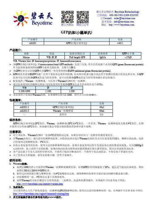
碧云天生物技术/Beyotime Biotechnology 订货热线:400-168-3301或800-8283301 订货e-mail :****************** 技术咨询:***************** 网址:碧云天网站 微信公众号GFP 抗体(小鼠单抗)产品编号 产品名称包装 AG281GFP 抗体(小鼠单抗)>40次产品简介:来源 用途 抗体识别位点抗体类型 GFP 分子量MouseWB, IP, IFFull length GFP IgG2a~27kDWB, Western blot; IP, Immunoprecipitation; IF, Immunofluorescence. 本GFP 抗体(小鼠单抗),即mouse monoclonal GFP antibody ,为进口分装,用全长的来源于水母的GFP (green fluorescent protein)作为抗原制备得到的抗GFP 小鼠单克隆抗体。
克隆号为B-2。
本GFP 抗体可以识别GFP以及GFP 的一些突变体例如EGFP (enhanced green fluorescent protein)。
GFP 或其突变体EGFP 等被广泛用于基因表达效率的检测,以及和目的蛋白融合表达用于检测目的蛋白的表达和分布。
本GFP 抗体不仅可以检测GFP 或其适当的突变体,也可以检测和GFP 或其适当的突变体融合表达的蛋白。
配套提供了Western 一抗稀释液,可以用于Western 检测时的一抗稀释。
):40次。
包装清单:产品编号 产品名称包装 AG281-1 GFP 抗体(小鼠单抗) 40µl AG281-2 Western 一抗稀释液40ml —说明书1份保存条件:GFP 抗体(小鼠单抗)-20ºC 保存,Western 一抗稀释液-20ºC 或4ºC 保存,一年有效。

外泌体(exosomes)是由多种细胞(上皮细胞、巨噬细胞、间充质干细胞、免疫细胞等)主动向胞外分泌的一种纳米级别的胞外囊泡,在血液、尿液、羊水、脑脊液、淋巴液、母乳、唾液、胃酸等生物液中广泛分布[1]。
其最初被认知为细胞释放的代谢物,但后续的研究发现外泌体是细胞通信的重要介质[2],其携带的蛋白质、核酸、脂质等多种生物活性成分在细胞增殖、细胞凋亡、血管新生、免疫反应及细胞通信等众多生物学过程发挥重要作用[3]。
肺纤维化(pulmonary fibrosis,PF)是一种慢性、间质性的肺组织间质损伤疾病,也是多种肺部疾病的最终表现[4]。
其发病机制复杂,患者生存周期短、难治愈、病死率高,目前该疾病尚缺乏确切有效的治疗方法。
遗传异常、自身免疫、机体损伤等自身条件因素以及病原微生物感染、放射性元素、职业条件等外部环境因素都会引起复杂肺间质损伤,根据发病原因的不同,肺纤维化可分为特发性肺纤维化、继发性肺纤维化、遗传性肺纤维化以DOI:10.16605/ki.1007-7847.2022.04.0131收稿日期:2022-04-02;修回日期:2022-08-03;网络首发日期:2022-09-30基金项目:宁夏自然科学基金项目(NZ1633);国家自然科学基金资助项目(31660255)作者简介:王泽华(1997—),男,陕西渭南人,硕士研究生:*通信作者:马春燕(1975—),女,宁夏银川人,博士,教授,主要从事外泌体及动物病原微生物研究,E-mail:********************。
外泌体miRNA 及其在肺纤维化中的作用研究进展王泽华,张丽昀,张浩澜,胡成博,马春燕*(宁夏大学生命科学学院西部特色生物资源保护与利用教育部重点实验室,中国宁夏银川750021)摘要:外泌体是由细胞膜与多囊泡体融合并通过胞吐作用释放到胞外的纳米尺寸的脂双层囊泡,在细胞间通信作为媒介发挥重要作用。
微RNA (microRNA,miRNA)是一类由18~24个核苷酸组成的内源性非编码RNA,在转录后基因表达中充当重要的调节因子。


Potential autocrine regulation of interleukin-33/ST2signaling of dendritic cells in allergic inflammationZ Su 1,2,J Lin 2,3,F Lu 1,X Zhang 1,2,L Zhang 2,3,NB Gandhi 2,CS de Paiva 2,SC Pflugfelder 2and D-Q Li 2This study identified a novel phenomenon that dendritic cells (DCs)produced interleukin (IL)-33via Toll-like receptor (TLR)-mediated innate pathway.Mouse bone marrow–derived DCs were treated with or without microbial pathogens or recombinant murine IL-33.IL-33mRNA and protein were found to be expressed by DCs and largely induced by several microbial pathogens,highly by lipopolysaccharide (LPS)and ing two mouse models of topical challenge by LPS and flagellin and experimental allergic conjunctivitis,IL-33-producing DCs were observed in ocular mucosal surface and the draining cervical lymph nodes in vivo .The increased expression levels of myeloid differentiation primary-response protein 88(MyD88),nuclear factor (NF)-j B1,NF-j B2,and RelA accompanied by NF-j B p65nuclear translocation were observed in DCs exposed to flagellin.IL-33induction by flagellin was significantly blocked by TLR5antibody or NF-j B inhibitor quinazoline and diminished in DCs from MyD88knockout mice.IL-33stimulated the expression of DC maturation markers,CD40and CD80,and proallergic cytokines and chemokines,OX40L,IL-4,IL-5,IL-13,CCL17(C-C motif chemokine ligand 17),TNF-a (tumor necrosis factor-a ),and IL-1b .This stimulatory effect of IL-33in DCs was significantly blocked by ST2antibody or soluble ST2.Our findings demonstrate that DCs produce IL-33via TLR/NF-j B signaling pathways,suggesting a molecular mechanism by which local allergic inflammatory response may be amplified by DC-produced IL-33through potential autocrine regulation.INTRODUCTIONInterleukin-33(IL-33),a new member of the IL-1super family,has been recently identified as a functional ligand to ST2.By binding to ST2receptor,IL-33activates T helper type 2(Th2)cells and mast cells to secrete Th2cell–associated proin-flammatory cytokines and chemokines that lead to allergic pathological changes in mucosal organs.1More and more evidences show that IL-33has an important role in inflam-matory diseases:hypersensitive diseases like asthma,2,3auto-immune diseases like rheumatoid arthritis,4dermatitis,5,6colitis,7–9allergic rhinitis,10and conjunctivitis.11,12Major sources of IL-33expression include epithelial and endothelial cells,as well as fibroblasts and others.1,2,13–15Until recently,Yanagawa and colleagues reported that murine bone marrow–derived dendritic cells (DCs)could express IL-33.16,17DCs are the most potent professional antigen-presenting cells linking innate and adaptive immune response.DCs express a variety of Toll-like receptors (TLRs),which recognizeconserved microbial components and have an important role in the mucosal innate immune system.18Mucosal surfaces contain resident DCs capable of sensing the external stimuli and mounting local responses upon recognition of invading microorganisms.19We have demonstrated that IL-33expres-sion is an innate response to certain microbial pathogens through TLRs and nuclear factor (NF)-k B signaling pathways in human corneal epithelial cells.20However,there is no report that DCs produce IL-33via TLR-mediated innate immunity signaling.As in most tissues,ocular surface DCs are present at very low concentrations and are difficult to isolate.21DCs induced from mouse bone marrow have been widely used for the study.22,23Using both in vitro cultured bone marrow–derived DCs and in vivo ocular surface of BALB/c mice topically challenged with microbial pathogens,the present study explored a novel phenomenon that DCs produce IL-33via TLR-mediated innate signaling pathway in response to microbial pathogens,which may have an important role in1School of Optometry and Ophthalmology,Wenzhou Medical College,Wenzhou,China.2Ocular Surface Center,Cullen Eye Institute,Department of Ophthalmology,Baylor College of Medicine,Houston,Texas,USA and 3Department of Ophthalmology,the Affiliated Hospital of Qingdao University Medical College,Qingdao,China.Correspondence:F Lu (lufan@)or D-Q Li (dequanl@)Received 26July 2012;accepted 13November 2012;published online 9January 2013.doi:10.1038/mi.2012.130nature publishing groupARTICLESSee ARTICLE page 911amplifying the allergic inflammatory response in local mucosa through a potential autocrine regulatory mechanism.RESULTSIL-33was produced by mouse DCs in response to specific TLR ligandsMouse DCs were induced from bone marrow cells by GM-CSF (granulocyte macrophages colony-stimulating factor)in cul-ture for 9days,and the purity of DCs was 492%,as evaluated by DC surface marker CD11c with flow cytometry analysis.The DCs were treated with or without extracted or synthetic microbial products that are ligands to TLRs 1–9(10m g ml À1of Pam3CSK4,peptidoglycan (PGN),flagellin,diacylated lipo-protein (FSL-1),R837,single-stranded RNA (ssRNA),type C CpG oligonucleotide (C-CpG-ODN),1m g ml À1of lipopoly-saccharide (LPS),or 50m g ml À1of Polyinosinicpolycytidylic acid (polyI:C))for 4–24h.IL-33mRNA was found to be expressed at very low level in untreated DCs,but it was largely induced by specific TLR ligands (Figure 1a ).The induction of IL-33mRNA expression in DCs reached the peak level at 8h (Figure 1b ).As shown in Figure 1a,b ,LPS and flagellin significantly induced IL-33mRNA expression to 6–10-fold (P o 0.01)in a dose-dependent fashion;Pam3CSK4and polyI:C also upregulated IL-33mRNA by 3–4-fold (P o 0.05);whereas PGN,FSL-1,R837,ssRNA,and C-CpG-ODN did not significantly induce IL-33expression.To confirm the IL-33production at protein level,DCs were exposed to LPS and flagellin for 24h,which were microbial ligands to TLRs 4and 5,respectively,and strongly stimulated the IL-33protein expression.IL-33protein was barely detected in the cell lysates from untreated DCs but was significantly stimulated to 2–3-fold by LPS and flagellin (P o 0.05),as determined by enzyme-linked immu-nosorbent assay (ELISA;Figure 1c )and western blotting (Figure 1d ).The immunofluorescent staining further showed that IL-33was immunolocalized in the nucleus and cytoplasma in normal DCs,and IL-33-positive cells were largely increased with more significant cytoplasmic immunostaining in DCs treated with LPS or flagellin (Figure 1e ).Furthermore,we observed that LPS and flagellin significantly upregulated DC maturation markers,CD40,CD80,CD86,and MHC (major histocompatibility complex)class II,with more and large clumps formed in DC cultures,indicating that DC maturation may contribute to IL-33induction by microbial ligands (see Supplementary Figure S1online).Figure 1Dendritic cells (DCs)produce interleukin (IL)-33in response to microbial pathogens.(a )IL-33mRNA expression was determined by quantitative real-time PCR in murine DCs from BALB/c mice exposed to microbial products,ligands to Toll-like receptors 1–9(10m g ml À1of Pam3CSK4,peptidoglycan (PGN),flagellin,diacylated lipoprotein (FSL-1),R837,single-stranded RNA,type C CpG oligonucleotide (C-CpG-ODN),1m g ml À1of lipopolysaccharide (LPS)or 50m g ml À1of polyinosinicpolycytidylic acid (polyI:C))for 8h.(b )The time course and dose response of IL-33mRNA by DCs exposed to LPS or flagellin.(c ,d )IL-33protein production in cell lysates of DCs treated with LPS (1m g ml À1)or flagellin (10m g ml À1)for 24h was determined by enzyme-linked immunosorbent assay and western blotting,respectively.(e )Representative images showing IL-33immunoreactivity (green)in DCs exposed to LPS (1m g ml À1)or flagellin (10m g ml À1)for 24h by immunofluorescent staining (Green)with propidium iodide (Red)as nuclear counterstaining.Each bar in the diagrams represents mean ±s.d.of three to five independent experiments.*P o 0.05;**P o 0.01.MW,molecular weight.The color reproduction of this figure is available on Mucosal Immunology journal online.ARTICLESIL-33was produced by CD11c þDCs infiltrated inconjunctiva and migrated to cervical lymph nodes (CLNs)of mice topically challenged by LPS or flagellinTo further identify whether DCs produce IL-33in vivo ,LPS (1m g per 5m l phosphate-buffered saline (PBS)per eye)orFigure 2Mucosal dendritic cells (DCs)produce interleukin (IL)-33in murine conjunctiva (Conj)and cervical lymph nodes (CLN)in vivo .BALB/c mice were topically challenged in the conjunctival sac with 1m g lipopolysaccharide (LPS)or flagellin in 5m l phosphate-buffered saline (PBS)per eye,three times a day for 2days,5m l PBS was used as a control.Frozen sections of eyeballs ((a )showing cornea and conjunctiva)and (b )CLN were used for double immunofluorescent staining with CD11c (Red)and IL-33(Green)using DAPI (40,6-diamidino-2-phenylindole)counterstaining (Blue).Arrows indicate positive staining signals.Each bar in the diagrams represents mean ±s.d.of three independent experiments.*P o 0.05;**P o0.01.Figure 3Interleukin (IL)-33-producing dendritic cells in murineexperimental allergic conjunctivitis (EAC).The EAC model was induced in BALB/c mice sensitized and topically challenged by SRW pollen (EAC),with phosphate-buffered saline (PBS)-treated mice (PBS)as controls.Frozen sections of eyeballs ((a )showing cornea and conjunctiva)and (b )cervical lymph nodes (CLNs)were used for double immunofluorescent staining with CD11c (Red)and IL-33(Green)using DAPI (40,6-diamidino-2-phenylindole)counterstaining (Blue).Arrows indicate positive staining signals.Each bar in the diagrams represents mean ±s.d.of three independent experiments.*P o 0.05;**P o 0.01.ARTICLESflagellin(1m g per5m l PBS per eye)was topically instilled in the conjunctival sac of BALB/c mice three times a day for2 days.The mice treated with5m l PBS alone were used as controls.As shown in Figure2,the double immunofluorecent staining revealed that murine corneal and conjunctival epithelia weakly expressed IL-33in normal control mice but produced strong IL-33immunoreactivity when stimulated by LPS or flagellin,indicating that IL-33is mainly produced by epithelial cells,a similar pattern to human ocular surface epithelia.20Interestingly,IL-33immunoreactivity was also observed in CD11cþDCs that infiltrated into the conjunctival stroma near epithelial area(Figure2a)and draining CLNs (Figure2b)in the mice challenged with LPS or flagellin.This finding further identified that DCs produce IL-33in vivo in murine mucosal ocular surface and draining CLNs in response to microbial pathogens.IL-33-producing DCs in murine experimental allergic conjunctivitis(EAC)To confirm the role of IL-33-producing DCs in allergic disease, EAC model was induced in BALB/c mice sensitized and topically challenged by short ragweed(SRW)pollen(EAC mice),with PBS treated(PBS mice)as control groups. Repeated topical challenges with SRW allergen generated typical signs mimic to human allergic conjunctivitis,including lid edema,conjunctival redness,chemosis,tearing,and fre-quent scratching of the eye lids.Consistent with our previous reports,24,25the infiltration of CD11cþDCs on the ocular surface was detected in this EAC model by immunostaining.As shown in Figure3,CD11cþDCs were accumulated in the SRW-challenged ocular surface,primarily in the stroma subjacent to conjunctival epithelia.Interestingly,double staining showed some DCs producing IL-33(Figure3a).In draining CLN,we also observed the dramatic increase of positively immunoreactive cells to both CD11c and IL-33 (Figure3b).These results suggest a potential role of IL-33-expressing DCs in EAC mice.IL-33induction was mediated via TLR/NF-j B signaling pathways by flagellinMyeloid differentiation primary-response protein88(MyD88) is a universal adapter protein necessary for response to most TLRs except TLR3.26,27TLR signaling typically induces activation of the NF-k B.Therefore,we hypothesized that LPS and flagellin promoted IL-33production via TLR/NF-k B signaling pathways.Taking flagellin as an example,we observed the significant increase of MyD88,NF-k B1,and NF-k B2(all P o0.01),as well as RelA(P o0.05)that encodes NF-k B p65,at mRNA levels,in the DCs exposed to flagellin(Figure4a). Immunofluorescent staining revealed that NF-k B p65protein was mainly located in cytoplasm in untreated control DCs but was markedly translocalized from cytoplasm to nucleus in DCs exposed to flagellin(Figure4b),indicating NF-k B signaling activation.Interestingly,the stimulated mRNA expression of MyD88,NF-k B1,and NF-k B2,the nuclear translocation of NF-k B p65,and the increased IL-33production by flagellin were significantly blocked by TLR5antibody or quinazoline,a NF-k B activation inhibitor(NF-k B-I),with an exception that NF-k B-I did not block the stimulated MyD88mRNA,an upstream molecule of NF-k B(Figure4). Furthermore,we cultured bone marrow–derived DCs from MyD88À/Àmice and their age-and gender-matched wild-type MyD88þ/þlittermates to investigate whether MyD88signal-ing is essential for TLR activation and IL-33induction.As shown in Figure5a,the mRNA expression levels of NF-k B signaling(NF-k B1,NF-k B2,RelA)and IL-33induction (P o0.01,0.01,0.05and0.01,respectively)were strongly stimulated by flagellin in DCs derived from MyD88þ/þmice when compared with the untreated control.But these stimulatory effects of flagellin were largely abolished in DCs derived from MyD88À/Àmice.IL-33induction,evaluated by its immunoreactivity(Figure5b),was also significantly increased by flagellin in DCs of MyD88þ/þwild type but not MyD88À/Àmice.Potential autocrine regulation of IL-33/ST2in DCs activationDCs have an important role in initiating and maintaining the Th2-dominant allergic inflammation.28IL-33has been identi-fied as the ligand of ST2,the well-known receptor in Th2cells.1 Through binding to ST2,IL-33promotes Th2cells producing Th2inflammatory cytokines IL-4,IL-5,and IL-13in allergic disease.Based on a recent report that ST2was identified to be expressed by DCs,23our finding that IL-33is produced by DCs in response to microbial pathogens leads us to hypothesize that DCs may amplify allergic inflammatory response via potential autocrine regulation in IL-33/ST2signaling.Mouse DCs were incubated with1ng mlÀ1of recombinant murine(rm)IL-33for4or24h.As evaluated by quantitative real-time PCR(RT-qPCR)and flow cytometry,we found that IL-33increased the expression of CD40and CD80at both mRNA and protein levels(P o0.05;Figure6a,b),suggesting that IL-33may have a role in promoting DC maturation.We have observed that IL-33activated DCs to produce Th2 cytokines(IL-4,IL-5,and IL-13),Th2-attracting chemokine CCL17(C-C motif chemokine ligand17),and proinflammatory cytokines(TNF-a(tumor necrosis factor-a)and IL-1b;Figure6a).More interestingly,we observed a previously unknown phenomenon that IL-33primes DCs to produce Th2-inducing cytokine OX40ligand(OX40L)at both mRNA and protein levels(Figure6a,b),which is consistent with a recent study.29OX40L is well known to be induced in activated DCs by thymic stromal lymphopoietin,an epithelial-derived proallergic cytokine,to trigger Th2-dominant allergic inflammatory response.Interestingly,the stimulated expression of DC maturation markers(CD40and CD80) and proallergic cytokines and chemokines(OX40L,IL-4, IL-5,IL-13,CCL17,TNF-a,and IL-1b)by IL-33was significantly blocked by ST2neutralizing antibody or exogenous soluble ST2protein.These results demonstrate that DCs may have an important role in amplifying local allergic inflammation through a potential autocrine regulation of IL-33/ST2signaling.ARTICLESDISCUSSIONIL-33has been consolidated as a pro-inflammatory mediator in allergic inflammation.1,30–32IL-33signals through a hetero-dimeric membrane receptor composed of ST2and IL-1R1accessory protein and activates Th2lymphocytes,mast cells,eosinophils,basophils,macrophages,NK (natural killer),and NKT cells.33,34The environmental or endogenous triggers that provoke IL-33cellular release may be associated with infection,inflammation,or tissue damage.2Relatively abundant IL-33mRNA expression is found in multiple tissue–related cell types such as mucosal epithelial cells,endothelial cells,fibroblasts,smooth muscle,cardiomyocytes,keratinocytes,and adipo-cytes.1,13,35Skin,gut,and lung appear to be prominent IL-33-expressing organs.However,whether DCs express IL-33is not clear,although one group has reported recently that DCs produce IL-33during inflammation.16,17The involvement of bacterial agents like LPS and flagellin in initiation of allergicinflammation has been recently documented.36–41In this study,we demonstrated that IL-33was induced by mouse DCs,possibly via TLR/NF-k B signaling pathways in response to microbial pathogens.The DC-produced IL-33may amplify local inflammatory response through autocrine mechanism.DCs produce IL-33both in vitro and in vivo in response to microbial pathogensBased on the important role of DCs in innate immunity and the observation that IL-33is mainly produced by epithelial cells via TLR-mediated innate response,20we hypothesized that DCs are capable of producing IL-33in response to microbial pathogens.We incubated the murine bone marrow–derived DCs with TLR ligands 1–9and found that several TLR ligands,especially LPS and flagellin,the ligands to TLR4and TLR5,respectively,significantly stimulated IL-33expression by mouse DCs at both the mRNA (P o 0.01)and protein levels (P o 0.05),asFigure 4Interleukin (IL)-33is induced in dendritic cells (DCs)via Toll-like receptor (TLR)/nuclear factor (NF)-k B signaling pathways.(a )The mRNA expression levels of MyD88(myeloid differentiation primary-response protein 88),NF-k B1,NF-k B2,RelA,and IL-33were evaluated by quantitative real-time PCR in DCs from BALB/c mice treated with flagellin (10m g ml À1)with or without TLR5antibody or quinazoline (NF-k B-I)1h preincubation for 8h.(b )Representative images showing NF-k B p65nuclear translocation and IL-33production by immunofluorescent staining (Green)with propidium iodide counterstaining (Red)in DCs treated as described in a for 24h.*P o 0.05;**P o 0.01,compared with control;^P o 0.05;^^P o 0.01,compared with flagellin.The color reproduction of this figure is available on Mucosal Immunology journal online.ARTICLESdetermined by RT-qPCR,ELISA,western blotting,and immunofluorescent staining (Figure 1).DCs are highly mobile and are present in the right place at the right time for the regulation of immunity.They are positioned as sentinels in the periphery,where they frequently encounter foreign antigens and penetrate epithelium to sample antigens,19then they readily relocate to secondary lymphoid organs,particu-larly lymph nodes,to position themselves optimally for encounter with naive or central memory T cells.42,43Using a topical challenge mouse model with LPS and flagellin,we further identified that DCs produce IL-33in vivo .The infiltrated CD11c þDCs that produce IL-33were observed in the conjunctiva of LPS-or flagellin-challenged eyes,as evaluated by immunofluorescent staining (Figure 2).Interestingly,we further found that CD11c þDCs migrated to CLNs and expressed high level of IL-33.The double-reactive cells (IL-33þCD11c þ),the most should be DCs,in CLN significantly increased 3.7–4.5-fold,from 4.14±1.26%in control mice to 18.64±3.05and 15.28±2.52%in LPS-or flagellin-challenged mice,respectively.Furthermore,IL-33-producing DCs were observed to accumulate in the ocular surface and the draining lymph nodes in a murine EAC model,as evaluated by double staining with CD11c and IL-33antibodies (Figure 3).The double-reactive cells (IL-33þCD11c þ)in CLN significantly increased 5.2-fold,from 3.87±0.61%in PBS control mice to 20.24±3.12%in EAC mice.DCs produce IL-33via TLR/NF-j B signaling pathwaysLPS-and flagellin-induced inflammation is mediated by TLR4and TLR5,respectively.44,45TLRs,which recognize conserved microbial components,are important pattern-recognition receptors.TLRs consist of a family of at least 11mammalian receptors that bind a restricted repertoire of ligands and recruit common adapter molecules to induce cell signaling.46MyD88is a universal adapter protein necessary for response to most TLRs,including TLR4and TLR5.26,27TLR signaling typically induces activation of NF-k B.NF-k B1or NF-k B2is bound to RelA to form the NF-k B complex.Transcription factor p65is encoded by the RelA gene.47Activated NF-k B complex translocates into the nucleus and binds DNA at kappa-B-binding ing flagellin as a model,we observed that flagellin significantly increased the expression of MyD88,NF-k B1,NF-k B2,RelA,and IL-33,as well as NF-k B activation with p65nuclear translocation (Figure 4).The stimulated IL-33induction by flagellin was markedly blocked by TLR5antibody and NF-k B inhibitor.Furthermore,we observed that the IL-33induction by flagellin was significantly reduced in DCs derived from MyD88knockout mice when compared with that seen in their wild-type littermates.All these results demonstrated that IL-33production in DCs was via TLR/NF-k B signaling pathways in response to microbial pathogens.DCs may amplify local inflammatory response through a potential autocrine mechanismDCs have an important role in initiating and maintaining an allergic Th2immune response.28Mucosal epithelium–derived IL-33can activate Th2cells and mast cells to produce Th2inflammatory cytokines and chemokines through binding to ST2receptor.1As DCs could express ST2,23our finding that DCs also produce IL-33suggest that DCs may amplify inflammatory response via potential autocrine mechanism.In this study,we observed that exogenous IL-33stimulated expression of costimulatory molecules CD40,CD80,and OX40L,as well as Th2inflammatory cytokines and chemokines in DCs.Furthermore,these stimulatory effects of IL-33were significantly blocked by ST2antibody or soluble ST2protein.The role of IL-33-producing DCs was further observed in an EAC mouse model (Figure 3).DC-produced IL-33may serve as a source of IL-33for other immune cells expressing ST2,potentially amplifying allergic inflammation through a paracrine mechanism.33,34In summary,this study revealed that microbial pathogens,like LPS and flagellin,induce expression and production of pro-allergic cytokine IL-33by DCs via TLR/NF-k B signaling pathways.DCs not only respond to IL-33but also produce IL-33in allergic condition.Our findings suggest a novel mechanism by which local inflammatory response may be amplified by DC-produced IL-33through a potential autocrine mechanism,which may provide a therapeutic potential to treat ocular allergic disease through a local blockade of IL-33produced byDCs.Figure 5Interleukin (IL)-33is induced in dendritic cells (DCs)via MyD88(myeloid differentiation primary-response protein 88)signaling pathways.(a )The mRNA expression of nuclear factor (NF)-k B1,NF-k B2,RelA,and IL-33was evaluated by quantitative real-time PCR in DCs from MyD88þ/þand MyD88À/Àmice exposed to flagellin (10m g ml À1)for 8h.(b )Representative images showing IL-33production by immunofluorescent staining (Green)with propidium iodidecounterstaining (Red)in DCs from MyD88þ/þand MyD88À/Àmice treated with flagellin (10m g ml À1)for 24h.Each bar in the diagrams represents mean ±s.d.of three independent experiments.*P o 0.05;**P o 0.01,compared with MyD88þ/þcontrol,^P o 0.05;^^P o 0.01,compared with MyD88þ/þflagellin.ARTICLESMETHODSMaterials and reagents .Cell culture dishes,plates,centrifuge tubes,and other plasticware were purchased from BD Biosciences (Lincoln Park,NJ);polyvinylidene difluoride (PVDF)membrane was from Millipore (Bedford,MA);polyacrylamide ready gels (4–15%Tris-HCl),sodium dodecyl sulfate (SDS),prestained SDS-poly-acrylamide gel electrophoresis low range standards,precision plus protein standards,and precision protein Strep-Tactin horseradish peroxidase conjugate were from Bio-Rad (Hercules,CA);RPMI-1640medium,amphotericin B and gentamicin were from Invitrogen (Grand Island,NY);fetal bovine serum from Hyclone (Logan,UT).The extracted or synthetic microbial components,Pam3CSK4,PGN from Bacillus subtilis (PGN-BS),flagellin from Salmonella typhi-murium ,FSL-1,imiquimod (R837),single-stranded,GU-richoligonucleotide complexed with LyoVec (ssRNA40/LyoVec),and type C CpG oligonucleotide (ODN 2395)were from invivoGen (Santiago,CA).polyI:C and LPS from Escherichia coli were from Sigma-Aldrich (St Louis,MO).rmGM-CSF,rmIL-4,and rmIL-33were from Peprotech (Rochy Hill,NJ).Rabbit polyclonal IL-33antibody was from Santa Cruz Biotechnology (Santa Cruz,CA).Rabbit polyclonal ST2antibody was from Enzo (Farmigdale,NY).rmIL-33and rmST2soluble proteins were from R&D (Minneapolis,MN).Hamster monoclonal CD11c antibody was from Abcam (Cambrige,MA).Mouse IL-33ELISA kit,purified anti-mouse NF-k B p65,purified anti-mouse CD16/32,fluorescein isothiocyanate–conjugated anti-mouse CD11c,allophycocyanin (APC)-conjugated anti-mouse CD40,APC-conjugated anti-mouse CD80,phycoerythrin(PE)-conjugated anti-CD86,and PE-conjugated anti-mouse I-A/I-E were fromBiolegendFigure 6Interleukin (IL)-33activates dendritic cells (DCs)in vitro .(a )The expression of cytokines and chemokines was evaluated byquantitative real-time PCR in DCs from BALB/c mice exposed to IL-33(1ng ml À1)for 4h without or with 1h previous incubation of ST2antibody (Ab)or soluble ST2protein.(b )Flow cytometry showed the enhanced expression of CD40,CD80,and OX40L of DCs exposed to IL-33for 24h.Each bar in the diagrams represents mean ±s.d.of three independent experiments.*P o 0.05;**P o 0.01,***P o 0.001,compared with control;^P o 0.05,compared with IL-33.APC,allophycocyanin;CCL,C-C motif chemokine ligand;FITC,fluorescein isothiocyanate;MFI,mean fluorescent intensity;PE,phycoerythrin;SSC-A,side scatter area;TNF,tumor necrosis factor.ARTICLES(San Diego,CA).RNeasy Mini RNA extraction kit was from Qiagen (Valencia,CA);enhanced chemiluminescence reagents and Ready-To-Go-Primer First-Strand Beads were obtained from GE Healthcare (Piscataway,NJ);TaqMan gene expression assays and real-time PCR master mix were from Applied Biosystems(Foster City,CA);and horseradish peroxidase–conjugated goat anti-rabbit immunoglobulin G and BCA(bicinchoninic acid)protein assay kit were from Pierce Chemical(Rockford,IL).Animal.The animal research protocol was approved by the Center for Comparative Medicine at Baylor College of Medicine.All animals used in this study were maintained in specific pathogen-free conditions in microisolator cages and were treated in accordance with the guidelines provided in the Association for Research in Vision and Ophthal-mology statement for the use of animals in ophthalmic and vision research.Female BALB/c mice at6–8-weeks old were purchased from the Jackson Laboratory(Bar Harbor,ME).Heterozygous MyD88À/Àmice on a C57BL/6background were kindly provided by Dr Shizuo Akira(Research Institute for Microbial Disease,Osaka University, Japan)through Dr Eric Pearlman(Department of Ophthalmology and Visual Sciences,CaseWestern Reserve University,Cleveland,OH). The genotyping was performed by means of PCR of the tail DNA with three specific primers(MyD88F,TGGCATGCCTCCATCAT AGTTAACC;MyD88R,GTCAGAAACAACCACCACCATGC; MyD88R-Neo,ATCGCCTTCTATCGCCTTCTTGACG)by using a previously described method.48The MyD88À/Àmice grown to 6–8weeks were used for experiments.Generation of murine bone marrow–derived Dcs.Bone marrow–derived DCs were generated as previously described22with minor modifications.Briefly,6–8-week-old female BALB/c mice were killed, femurs and tibiae were removed,and the marrow was flushed with RPMI-1640using a syringe with a0.3-mm needle.Clusters within the marrow suspension were disassociated by vigorous pipetting.Bone marrow cells were suspended in complete media(CM,RPMI-1640 supplemented with10%fetal bovine serum,50m g mlÀ1gentamicin, and1.25m g mlÀ1amphotericin B).Cells were adjusted to1.0Â106mlÀ1and plated on100-mm dish at10ml per well.They were cultured in CM containing15ng mlÀ1of rmGM-CSF and5ng mlÀ1 of rmIL-4(CM-CSF-IL-4)at371C and5%CO2.On day3of culture, 10ml of CM-CSF-IL-4was added to the cells.On days6and8,10ml of medium with cells were collected from each culture dish and centri-fuged at300g for5min at room temperature;the cells were re-suspended in10ml of the same medium,and then given back to the dishes.On day9,the non-adherent DCs were used for experiments. Treatment of murine bone marrow-derived DCs.DCs at1.0Â106 per well in12-well plates were incubated for4–24h with medium alone,TLR ligands(Pam3CSK4,PGN,polyI:C,LPS,flagellin,FSL-1, R837,ssRNA,or C-CpG-ODN,ligands to TLR1–9respectively, 10m g mlÀ1each,except for50m g mlÀ1polyI:C and1m g mlÀ1LPS), or IL-33(1ng mlÀ1)at the absence or presence of ST2neutralizing antibody(5m g mlÀ1)or soluble ST2protein(5m g mlÀ1)for mRNA expression.DCs at1.0Â106or5.0Â106were treated with LPS, flagellin,or IL-33(1ng mlÀ1)for24h in500m l medium for protein analysis by ELISA,immunofluorescent staining,western blotting,or flow cytometry.A topical challenge murine model of ocular surface with LPS and flagellin.BALB/c mice were topically challenged in the conjunctival sac with1m g LPS or1m g flagellin in5m l PBS per eye three times a day for2days,5m l PBS alone was used as a control.Twenty-four hours after the final challenge,the whole globes and draining CLNs in each group(n¼4)were excised,embedded in optimal cutting temperature compound(VWR,Suwanee,Ga),and flash-frozen in liquid nitrogen. Sagittal8-mm cryosections from murine globes and CLNs were cut with a cryostat(HM500;Micron,Waldorf,Germany)and stored at À801C before use.A murine model of EAC induced by SRW pollen.The murine EAC model was induced using the previously reported methods.24,25In brief,mice were immunized with50m g of SRW pollen(Greer Laboratories,Lenoir,NC)in5mg of Imject Alum(Pierce Bio-technology)by footpad injection on day0.Allergic conjunctivitis was induced by repeated topical challenges of1.5mg of SRW pollen suspended in10ml of PBS(pH7.2)into each eye once a day from days 10–12.PBS eyedrop–treated SRW-sensitized and untreated mice were used as controls.On day13,24h after the last SRW challenge,the whole eyeballs and CLNs were harvested for immunofluorescent staining. RNA extraction,reverse transcription,and RT-qPCR.Total RNA was extracted with a RNeasy Micro Kit(Qiagen,Valencia,CA) according to the manufacturer’s instructions,quantified with a spectrophotometer(NanoDrop ND-1000;Thermo Scientific,Wil-mington,DE),and stored atÀ801C.The first-strand cDNA was synthesized by reverse transcription from1m g of total RNA using Ready-To-Go You-Prime First-Strand Beads as previously descri-bed.49,50Quantitative real-time PCR was performed(Mx3005P QPCR System;Stratagene,La Jolla,CA)with20m l reaction volume con-taining5m l of cDNA,1m l gene expression assay,and10m l gene expression master mix(TaqMan;Applied Biosystems,Foster City,CA).TaqMan gene expression assays were used:GAPDH (glyceraldehyde3-phosphate dehydrogenase;Mm99999915_g1), IL-33(Mm00505403_m1),MyD88(Mm00440338_m1),NF-k B1 (Mm00476361_m1),NF-k B2(Mm00479807_m1),RelA(Mm00501346_ m1),CD40(Mm00441891_m1),CD80(Mm00711660_m1),OX40L (Mm00437214_m1),IL-4(Mm00445259_m1),IL-5(Mm99999063_m1), IL-13(Mm00434204_m1),TNF-a(Mm99999068_m1),IL-1b(Mm004 34228_m1),and CCL17(Mm00516136_m1).The thermocycler para-meters were501C for2min and951C for10min,followed by40cycles of951C for15s and601C for1min.A nontemplate control was included to evaluate DNA contamination.The results were analyzed by the comparative threshold cycle(Ct)methodand normalized by GAPDHas an internal control.51ELISA.Double-sandwich ELISA for mouse IL-33was performed, according to the manufacturer’s protocol,to determine the IL-33 protein level in cell lysates with different treatment.Absorbance was read at a reference wavelength of450nm by a VERSAmax microplate reader(Molecular Devices,Sunnyvale,CA).Western blot analysis.Western blot analysis was performed with a previously reported method.51Briefly,the cell lysates(50m g per lane, measured by a BCA protein assay kit)were mixed with6ÂSDS reducing sample buffer and boiled for5min before loading.The proteins were separated on an SDS polyacrylamide gel and elec-tronically transferred to PVDF membranes.The membranes were blocked with5%nonfat milk in TTBS(50mM Tris(pH7.5),0.9% NaCl,and0.1%Tween-20)for1h at room temperature and incubated, first with primary antibodies against IL-33(1:100,2m g mlÀ1)or b-actin(1:500,1m g mlÀ1)overnight at41C,and then with horseradish peroxidase-conjugated secondary antibody for1h at room tem-perature.The signals were detected with enhanced chemiluminescence reagent using a Kodak image station2000R(Eastman Kodak,New Haven,CT).Immunofluorescent staining.Indirect immunostaining was per-formed according to our previously reported methods.52,53In brief, DCs were fixed in4%paraformaldehyde and permeabilized with0.2% Triton X-100in PBS at room temperature for10min,respectively,and frozen sections of the tissue were fixed with acetone atÀ301C for 5min.Primary rabbit anti-mouse IL-33(4m g mlÀ1),rabbit anti-mouse NF-k B p65(2m g mlÀ1),and hamster anti-mouse CD11c (10m g mlÀ1)were applied for2h.AlexaFluor488-conjugated or594-conjugated secondary antibodies(Invitrogen)were incubated for1 h at room temperature,propidium iodide or40,6-diamidino-2-phenylindole were used for nuclear counterstaining.SecondaryARTICLES。

miR-33表达与炎症反应的关系miR-33是一种生物学功能十分重要的微小RNA分子,在调节细胞胆固醇代谢和基因表达中起着重要的作用。
miR-33的异常表达在许多疾病中都被发现,包括心血管疾病、代谢性疾病和癌症等。
miR-33通过调节脂质代谢和炎症反应,影响了这些疾病的进展和发展。
然而,关于miR-33与炎症反应之间的关系,我们现在仍知之甚少。
炎症反应是机体对各种激素和损伤刺激物的复杂反应,旨在清除损伤、感染及异物等。
炎症反应引起细胞浸润和供氧,控制局部代谢和控制内部环境的平衡,从而保护整个微生物组。
然而,当炎症反应失控时,其会引起各种严重的疾病发生。
因此,了解miR-33在炎症反应中的作用是非常重要的。
近年来的研究表明,miR-33在炎症反应中起着重要作用。
miR-33可以通过控制炎症相关的转录因子和信号通路来调节炎症反应。
例如,miR-33可以抑制炎症性信号通路Toll 样受体4(TLR4)和核因子-kB(NF-kB)的活化。
同时,miR-33还可以在发炎细胞和非发炎细胞之间调节胆固醇代谢,从而抑制致炎基因的表达。
miR-33还可以通过调节肝细胞脂质代谢来控制炎症反应。
肝细胞脂质代谢异常会导致脂肪肝等疾病的发生。
miR-33可以调节肝细胞的胆固醇代谢途径,从而影响肝细胞脂质代谢和肝脏炎症反应。
最近,研究人员在心血管疾病中发现了miR-33的作用。
miR-33在心肌梗塞中具有保护作用,可以抑制炎症反应和细胞凋亡。
另外,miR-33在动脉粥样硬化动物模型中被发现可以增加炎症反应和脂肪的聚集,从而导致动脉粥样硬化和心血管疾病进展。
虽然miR-33在炎症反应中的作用已经得到了初步证实,但还有很多问题需要进一步研究。
例如,在不同的疾病中,miR-33的表达模式是如何变化的?miR-33如何调节不同类型的细胞,如炎性细胞和非炎性细胞的炎症反应?在实际情况下,miR-33作为治疗手段的潜力如何?总之,miR-33在炎症反应中发挥着重要的作用。

管理好自己的情绪对学生有多重要观后感Managing one's emotions is of great importance when it comes to students. As an educator, the ability to regulate and control one's emotions is crucial in creating apositive learning environment, fostering better teacher-student relationships, and promoting overall academic success.作为一名教育工作者,管理好自己的情绪对学生来说至关重要。
在创造积极的学习环境、促进良好师生关系以及提升学术成就方面,调节和控制自己的情绪能力非常关键。
Firstly, managing emotions allows educators to establish a positive learning environment. When teachers are able to effectively handle their own feelings such as frustration, stress, or anger, they create a safe and supportive atmosphere for students. This enables learners to feel more comfortable expressing themselves and taking risks in their academic pursuits.管理情绪使教育工作者能够建立积极的学习环境。
当教师能够有效地处理自己的情绪,如沮丧、压力或愤怒时,他们为学生创造了一个安全和支持的氛围。

2024年江西中考英语作文预测As we approach the 2024 Jiangxi Middle School Entrance Exam, students across the province are diligently preparing for one of the most important tests in their academic lives. The English essay component is particularly crucial, as it evaluates students’ language proficiency, critical thinking, and ability to express their thoughts coherently. In anticipation of the exam, I offer a prediction for a potential essay topic and a model essay to aid students in their preparation.In recent years, the issue of environmental protection has become increasingly prominent. With the rapid development of industry and urbanization, the natural environment has been significantly impacted, leading to a range of environmental problems. This essay will discuss the importance of environmental protection, the consequences of neglecting it,and the steps individuals and communities can take to preserve our planet.Environmental protection is crucial for maintaining the health and sustainability of our planet. The environment provides essential resources such as clean air, water, and fertile soil, which are necessary for all forms of life. By protecting the environment, we ensure that these resources remain available for future generations. Additionally, a healthy environment supports biodiversity, which is vital for ecosystem stability and resilience. Biodiversity contributes to everything from the pollination of crops to the regulation of the climate, making it indispensable for human survival and well-being.Neglecting environmental protection can lead to severe consequences that affect both the planet and human society. Pollution, one of the most immediate results of environmental neglect, contaminates air, water, and soil, posing serious healthrisks to humans and wildlife. For instance, air pollution can cause respiratory diseases, cardiovascular problems, and even cancer. Water pollution can lead to waterborne diseases and disrupt aquatic ecosystems, while soil contamination can affect food safety and agricultural productivity.Moreover, the depletion of natural resources due to unsustainable practices can lead to scarcity, increased competition for resources, and conflicts. Deforestation, overfishing, and the excessive extraction of minerals are examples of activities that deplete resources faster than they can be replenished. Climate change, driven by greenhouse gas emissions from human activities, is another significant consequence of environmental neglect. It leads to extreme weather events, rising sea levels, and changes in weather patterns, which can have devastating effects on communities and economies.Individuals and communities play a crucial role in environmental protection. Simple actions, when collectively adopted, can make a significant difference. One of the most effective ways to protect the environment is through the reduction of waste. By practicing the three R’s—reduce, reuse, and recycle—individuals can minimize the amount of waste that ends up in landfills and the environment. Reducing consumption, reusing items, and recycling materials help conserve natural resources and reduce pollution.Another important step is conserving energy. Using energy-efficient appliances, turning off lights and electronic devices when not in use, and opting for renewable energy sources like solar and wind power can significantly reduce greenhouse gas emissions. Additionally, adopting sustainable transportation methods, such as walking, cycling, carpooling, or using public transportation, can help decrease air pollution and carbon footprints.Communities can also engage in environmental conservation efforts by protecting natural habitats and biodiversity. Planting trees, restoring wetlands, and creating protected areas for wildlife are effective ways to enhance ecosystem health and resilience. Educational programs and community initiatives can raise awareness about environmental issues and inspire collective action.Environmental protection is a shared responsibility that requires the concerted efforts of individuals, communities, governments, and international organizations. By understanding the significance of environmental protection, recognizing the consequences of neglect, and taking proactive steps to preserve our planet, we can ensure a sustainable and healthy environment for current and future generations. As students prepare for the 2024 Jiangxi Middle School Entrance Exam, it is essential to remember that their actions and attitudes towards the environment today will shape the world they inherittomorrow. Let us all commit to protecting our environment and fostering a more sustainable future.。

生态效益英语Ecological Benefits: Preserving the Balance of NatureIn today's rapidly changing world, the importance of ecological benefits cannot be overstated. As we strive to meet the growing demands of our modern society, it is crucial that we recognize the vital role that a healthy ecosystem plays in sustaining our way of life. The ecological benefits that arise from the delicate balance of nature are multifaceted and far-reaching, impacting not only our immediate environment but also the long-term viability of our planet.At the heart of ecological benefits lies the intricate web of interdependence that exists within the natural world. Each component of an ecosystem, from the smallest microorganism to the largest predator, plays a crucial role in maintaining the overall equilibrium. When this balance is disrupted, the consequences can be severe and far-reaching. The loss of a single species can have a cascading effect, altering the dynamics of the entire ecosystem and ultimately compromising the resources and services that we as humans rely upon.One of the primary ecological benefits is the provision of clean air and water. The plants and trees that populate our forests and landscapes act as natural air filters, absorbing pollutants and releasing oxygen into the atmosphere. This process not only improves the quality of the air we breathe but also helps to mitigate the effects of climate change by reducing the concentration of greenhouse gases in the atmosphere. Similarly, the complex network of rivers, lakes, and aquifers that make up our water systems are essential for providing clean, potable water for human consumption, agricultural use, and industrial processes.Another significant ecological benefit is the regulation of the global climate. The Earth's ecosystems, particularly the vast expanses of forests and oceans, play a crucial role in the carbon cycle, absorbing and storing vast quantities of carbon dioxide. This process helps to regulate the Earth's temperature and prevent the potentially catastrophic effects of global warming. Additionally, the diverse range of plant and animal life within an ecosystem contributes to the regulation of local and regional weather patterns, influencing factors such as precipitation, temperature, and humidity.The ecological benefits of a healthy ecosystem also extend to the realm of human health and well-being. Many of the medicines and treatments that we rely upon are derived from natural sources, suchas plants, fungi, and microorganisms. The loss of biodiversity threatens the potential discovery of new and potentially life-saving compounds. Furthermore, the presence of green spaces and natural environments has been shown to have a positive impact on mental health, reducing stress and promoting overall well-being.Beyond the direct benefits to human health and the environment, the preservation of ecological balance also has significant economic implications. The services provided by healthy ecosystems, such as flood control, soil fertility, and pollination, are essential for sustaining agricultural productivity and maintaining the viability of various industries. The tourism and recreation industries, in particular, are heavily dependent on the preservation of natural landscapes and the diverse array of plant and animal life that inhabit them.Despite the overwhelming evidence of the ecological benefits that arise from a healthy ecosystem, the reality is that many of these systems are under threat. The relentless pursuit of economic growth and development has led to the widespread degradation of natural habitats, the depletion of natural resources, and the introduction of pollutants and invasive species that disrupt the delicate balance of nature. As a result, we are witnessing the decline of biodiversity, the erosion of ecosystem services, and the increasing vulnerability of human communities to the effects of environmental change.To address these pressing challenges, it is imperative that we adopt a holistic and collaborative approach to environmental stewardship. This includes the implementation of sustainable practices in all sectors of the economy, the promotion of renewable energy sources, the protection of critical habitats and endangered species, and the active engagement of communities in the decision-making process. By recognizing the intrinsic value of the natural world and the vital role it plays in sustaining our way of life, we can work towards a future where the ecological benefits of a healthy ecosystem are not only preserved but actively celebrated and cherished.In conclusion, the ecological benefits that arise from a balanced and thriving ecosystem are truly invaluable. From the provision of clean air and water to the regulation of the global climate and the promotion of human health and well-being, the natural world is a vital and irreplaceable resource that we must safeguard for generations to come. By embracing a more sustainable and eco-conscious approach to development and resource management, we can ensure that the delicate balance of nature is maintained, and the ecological benefits that it provides continue to enrich and sustain our lives.。

英语作文商业贿赂Business Bribery。
Business bribery refers to the act of giving or receiving something of value in order to influence the actions of an individual or organization in a commercial transaction. It is a widespread problem in many countries and industries, and it has serious consequences for both the businesses involved and the overall economy.The practice of business bribery is unethical and illegal, as it undermines the principles of fair competition and damages the trust and integrity of the business environment. It can take many forms, such as cash payments, lavish gifts, or other favors, and it is often used to secure contracts, gain access to resources, or obtain favorable treatment from government officials.One of the main reasons why business bribery is so prevalent is the desire for businesses to gain acompetitive advantage and secure lucrative deals. In many industries, the competition for contracts and resources is fierce, and companies may resort to bribery in order to gain an edge over their rivals. This can create a culture of corruption and dishonesty, where businesses feel pressured to engage in unethical behavior in order to stay afloat.Another factor that contributes to the prevalence of business bribery is the lack of effective enforcement and regulation. In many countries, the laws and regulations governing bribery are weak or poorly enforced, which creates an environment where businesses feel they can get away with corrupt practices. This not only harms the businesses involved, but it also damages the reputation of the country as a place to do business.The consequences of business bribery are far-reaching and damaging. For businesses, it can lead to legal and financial repercussions, as well as reputational damagethat can be difficult to repair. It can also result in a loss of trust and confidence from customers, investors, andother stakeholders, which can have a long-term impact on the success and sustainability of the business.In addition to the impact on individual businesses, business bribery also has broader economic and social consequences. It can hinder economic development by distorting market competition and deterring investment, and it can also contribute to social inequality and injustice by favoring certain individuals or groups over others. In the long run, it can erode the foundations of a healthy and prosperous society.In order to address the problem of business bribery, it is essential for governments, businesses, and other stakeholders to work together to create a culture of transparency, accountability, and ethical behavior. This can be achieved through the implementation of strong laws and regulations, effective enforcement mechanisms, and the promotion of ethical business practices. Businesses also have a responsibility to establish clear policies and procedures to prevent bribery, and to educate their employees about the importance of ethical conduct.Furthermore, businesses should strive to create a culture of integrity and honesty, where ethical behavior is valued and rewarded. This can help to build trust and confidence among employees, customers, and other stakeholders, and it can also contribute to the long-term success and sustainability of the business.In conclusion, business bribery is a serious problem that has far-reaching consequences for businesses, economies, and societies. It undermines the principles of fair competition and damages the trust and integrity of the business environment. In order to address this issue, it is essential for governments, businesses, and other stakeholders to work together to create a culture of transparency, accountability, and ethical behavior. By doing so, we can help to create a business environment that is fair, competitive, and sustainable for the long term.。

有关植被好处的英语作文Vegetation plays a crucial role in sustaining life on our planet. From the lush rainforests to the sprawling grasslands, the diverse array of plant life provides countless benefits that are essential for the well-being of our environment and the human population. In this essay, we will explore the multifaceted advantages of vegetation and why it is crucial to protect and preserve this invaluable resource.One of the primary benefits of vegetation is its role in maintaining a balanced ecosystem. Plants act as the foundation of the food chain, providing sustenance for a wide range of animal species. The intricate web of life that exists within natural habitats is largely dependent on the presence of diverse vegetation. This biodiversity not only supports the survival of countless species but also helps to regulate the delicate balance of our ecosystems.Moreover, vegetation plays a vital role in the carbon cycle, which is essential for regulating the Earth's climate. Plants absorb carbon dioxide, a greenhouse gas, during the process of photosynthesis and release oxygen, a crucial component of the air we breathe. Thisprocess helps to mitigate the effects of climate change by removing excess carbon dioxide from the atmosphere and storing it in their biomass. The preservation of large-scale vegetation, such as forests, is particularly crucial in this regard, as they act as natural carbon sinks, sequestering vast amounts of carbon and helping to maintain a stable climate.In addition to their role in the carbon cycle, plants also contribute to the regulation of the water cycle. Vegetation helps to prevent soil erosion by stabilizing the ground and slowing the flow of water. This, in turn, helps to replenish groundwater reserves and reduce the risk of flooding during heavy rainfall. Furthermore, the transpiration process, where plants release water vapor into the atmosphere, contributes to the formation of clouds and the distribution of precipitation, which is essential for maintaining a healthy water cycle.Vegetation also plays a crucial role in supporting human health and well-being. Many medicinal plants and herbs have been used for centuries to treat a wide range of ailments, and modern pharmaceutical research continues to uncover the therapeutic potential of various plant species. Additionally, exposure to natural environments with abundant vegetation has been shown to have positive effects on mental health, reducing stress, improving mood, and enhancing cognitive function.Beyond their direct health benefits, plants also contribute to the aesthetic and recreational value of our surroundings. Urban areas with well-maintained parks, gardens, and street trees not only improve the visual appeal of the environment but also provide opportunities for outdoor activities, leisure, and relaxation. This, in turn, can have a positive impact on the overall quality of life for residents and visitors alike.Furthermore, vegetation plays a crucial role in supporting sustainable agricultural practices. The roots of plants help to maintain soil health by preventing erosion, improving nutrient cycling, and promoting the growth of beneficial microorganisms. This, in turn, supports the production of healthy and nutrient-rich crops, which are essential for food security and human nutrition.In conclusion, the benefits of vegetation are multifaceted and far-reaching. From regulating the climate and supporting biodiversity to enhancing human health and well-being, the preservation and protection of plant life are crucial for the sustainability of our planet. As we face the challenges of environmental degradation and climate change, it is essential that we recognize the vital role of vegetation and take proactive steps to conserve and restore these invaluable natural resources. By doing so, we can ensure a healthier, more resilient, and more sustainable future for generations to come.。
综 述?3-磷酸甘油酸脱氢酶促进丝氨酸合成在肿瘤进展中的机制崔畅婉,孙峥嵘Themechanismof3-phosphoglyceratedehydrogenasepromotingserinesynthesisintumorprogressionCUIChangwan,SUNZhengrongDepartmentofBiobank,ShengjingHospitalAffiliatedtoChinaMedicalUniversity,LiaoningShenyang110001,China.【Abstract】Upregulationofserinebiosyntheticpathwayactivityisadistinctcommonfeatureofmanycancers.3-phosphoglyceratedehydrogenase(PHGDH),thefirstrate-limitingenzymeinthispathway,ishighlyexpressedinmelanoma,breastandkidneycancer.PHGDHplaysanimportantroleintumorcellproliferation,metastasisandinvasion.Glycolysisintermediateproduct3-glycericacidphosphateoxidizedtohydroxypropionicacidphosphateunderPHGDHaction,andfinallysynthesizedserine.Serineconvertedtoglycineandthenplaysanimportantroleinthesynthesisofnucleotides,s-adenosylmethionine(SAM)andreducedglutathione(GSH).PHGDHisexpectedtobeanewtargetfortumortherapy.【Keywords】3-phosphoglyceratedehydrogenase,cancer,glucolysisModernOncology2021,29(05):0885-0888【指示性摘要】丝氨酸生物合成途径活性的上调是许多癌症明显的共同特征。

抑制灵长类动物miR33可增强胆固醇逆转运郑芳【期刊名称】《临床检验杂志(电子版)》【年(卷),期】2012(000)001【摘要】尽管降低低密度脂蛋白胆固醇的药物已经十分普及,心血管病仍然是西方人健康的头号杀手。
目前心血管新药开发正致力于提升高密度脂蛋白胆固醇的水平,MicroRNA(miRNAs)是一类重要的转录后调控因子,有证据表明miRNA可参与脂质代谢的调控,因而有望成为较理想的药物。
MiR33a和MiR33b的编码序列分别位于SREBF2和SREBF1基因的内含子区域,他们可以抑制ABCA1基因的表达,而ABCA1是一个重要的HDL合成调节因子。
近来的研究表明:用寡核苷酸类似物拮抗小鼠的MiR33a可有效提升血浆HDL水平,同时对抗动脉粥样硬化。
但是小鼠只有MiR33a,没有MiR33b,MiR33b只存在于大型哺乳动物的SREBF1基因。
因此,本文以非洲绿猴为研究对象,发现拮抗MiR33a和MiR33b可提高其肝细胞的ABCA1表达水平,并持续提升血浆HDL水平超过12周。
值得注意的是,miR-33的抑制还可以上调一系列脂肪酸氧化相关基因(CROT,CPT1A,HADHB和PRKAA1),下调参与脂肪酸合成的基因(SREBF1,F.ASN,ACLY和ACACA),继而大幅降低血浆VLDL相关的三酰甘油水平,这些发现是在小鼠研究中没有过的。
这些研究结果表明灵长类动物是一个极有效的药理模型,抑制MiR33a和MiR33b是一种可靠的提升HDL,降低VLDL的治疗手段,可用于心血管病相关的血脂异常靶向治疗。
【总页数】5页(P40-44)【作者】郑芳【作者单位】武汉大学中南医院检验科【正文语种】中文【中图分类】R341【相关文献】1.新的CCR5疫苗策略可抑制SHIV感染灵长类动物 [J],2.二甲双胍介导胆固醇逆转运抑制动脉粥样硬化 [J], 王夏蕾; 杨景达; 魏伟; 沈菊连; 陆璐; 吕昕儒; 肖建平; 薛偕华3.BET蛋白抑制剂JQ1增强索拉非尼对肝癌细胞的增殖抑制研究 [J], 王宇;范璐璐4.温胆汤通过调节胆固醇逆转运相关膜蛋白表达抑制ApoE-/-小鼠动脉粥样硬化斑块形成 [J], 陈玄晶;吴炳鑫;吴焕林;徐丹苹5.抑制自噬可以增强七叶皂苷钠对肝细胞性肝癌的抑制作用 [J], 方超;周大臣;崔笑;魏昇;耿小平因版权原因,仅展示原文概要,查看原文内容请购买。
MicroRNAs (miRNAs) are small (22-nt) endogenous double-stranded RNAs that have emerged as posttranscriptional regulators of physiological processes (3). miRNAs bind to complementary target sites in the 3′ untranslated regions (3′UTRs) of mRNAs, causing translational repression and/or mRNA destabilization (3). A single miRNA can have multiple targets, potentially providing simultaneous regulation of the genes involved in a physiological pathway. We explored whether miRNAs contribute to the maintenance of cholesterol homeostasis.We undertook an unbiased genome-wide screen of miRNAs modulated by cellular cholesterol content. We identified a subset of 21 miRNAs differentially regulated in human macrophages by cholesterol depletion and cholesterol enrichment, including several whose predicted gene targets are involved in cholesterol uptake, transport, and efflux (table S1).Confirmation of these candidates identified miRNAs that were both up- and down-regulated by cellular cholesterol content (fig. S1A). Sequence alignment revealed that one of these candidates, hsa-miR-33a and its mouse homolog mmu-miR-33 (referred to here as miR-33),is encoded within intron 16 of SREBF2, a gene that encodes a key transcriptional regulator of cholesterol uptake and synthesis (Fig. 1A) (1). Furthermore, the pre-miRNA is highly conserved in mammals (Fig. 1A), prompting us to select miR-33 for further characterization of its role in cholesterol metabolism.We found that in mouse peritoneal macrophages, miR-33 and SREBF2 expression were coordinately down-regulated by cholesterol loading, suggesting that these gene regulatory elements are cotranscribed (Fig. 1B). Furthermore, macrophages depleted of cholesterol with the 3-hydroxy-3-methylglutaryl coenzyme A (HMG-CoA) reductase inhibitor simvastatin showed robust up-regulation of both miR-33 and SREBF2, but not SREBF1(Fig. 1B). Levels of miR-33 were inversely correlated with the expression of another cholesterol-responsive gene product, the cholesterol transporter ABCA1 (Fig. 1B). Analysis of the kinetics of miR-33 induction revealed a concomitant increase in miR-33 and SREBP2levels with simvastatin treatment, consistent with their coregulation (fig. S1B). We nextdetermined whether miR-33 is regulated under physiologic conditions by measuring itsexpression in mice fed a chow, rosuvastatin-supplemented, or high-fat diet. Consistent withour in vitro observations, hepatic miR-33 levels were inversely correlated with cholesterollevels and ABCA1 expression and positively correlated with SREBF2 mRNA levels,suggesting that miR-33 is regulated by dietary cholesterol in vivo (Fig. 1C and fig. S1, Cand D). miR-33 levels were also regulated in two mouse models of hypercholesterolemia:Ldlr −/− and Apoe −/− mice. Hepatic and peritoneal macrophage miR-33 levels were markedlyreduced in Ldlr −/− mice that were fed a high-fat diet (Fig. 1D). Similarly, miR-33 levels inperitoneal macrophages from hypercholesterolemic Apoe −/− mice correlated inversely withcellular cholesterol ester content and expression of ABCA1 (fig. S2A). We next examinedmiR-33 expression in mouse tissues and various cell lines. In addition to macrophages,miR-33 was highly expressed in mouse and human hepatic cells and to a lesser extent inendothelial cells (fig. S2B). Furthermore, miR-33 was widely expressed in mouse tissuesand was particularly abundant in the brain and liver (Fig. 1E).To gain insight into the function of miR-33, we analyzed its potential gene targets, usingseveral miRNA target prediction algorithms (3). We identified putative binding sites formouse miR-33 in the 3′UTR of several genes involved in cellular cholesterol mobilization,including ABCA1 and ABCG1 and the endolysosomal transport protein NPC1 (fig. S3),suggesting that miR-33 coordinates cholesterol homeostasis through these pathways (4, 5).To test this hypothesis, we determined the effect of miR-33 on the expression of ABCA1,ABCG1, and NPC1 in macrophages treated with acetylated low-density lipoprotein(AcLDL) (to enrich in cholesterol) or the LXR ligand T0901317 (to directly stimulateexpression of the three genes) (2). Transfection of mouse peritoneal macrophages withNIH-PA Author ManuscriptNIH-PA Author ManuscriptNIH-PA Author ManuscriptmiR-33 (but not a control miRNA) strongly decreased the stimulation of both ABCA1 and ABCG1 protein and mRNA (Fig. 2A and fig. S4A; quantification in fig. S5A). These effects of miR-33 were seen with concentrations of as little as 5 nM (Fig. 2B) and were reversible by co-incubation with an antisense inhibitor of miR-33 (anti-miR-33) (Fig. 2C;quantification in fig. S5, B and C). miR-33 also repressed ABCA1 and ABCG1 protein in mouse hepatic cells, indicating that its effects are not cell type–specific (Fig. 2D). Inhibition of endogenous miR-33 by anti-miR-33 increased the expression of ABCA1 and ABCG1 in macrophages and of ABCA1 in hepatocytes, which is consistent with the hypothesis that miR-33 has a physiological role in regulating the expression of these transporters (Fig. 2E;quantification in fig. S6).Although we found that miR-33 comparably repressed ABCA1 in cells of mouse and human origin, this was not the case for NPC1 and ABCG1. miR-33 strongly suppressed NPC1protein in cells of human origin (Fig. 2D), whereas in mouse cells, miR-33 suppressed NPC1 protein levels only modestly and had no effect on NPC1 mRNA levels (fig. S4A).Furthermore, transfection of human macrophage, hepatic, and endothelial cells with miR-33had no detectable effect on ABCG1 protein (Fig. 2D and fig. S4B). Conservation map analysis revealed that although the predicted miR-33 target sites in the 3′UTR of mouse ABCA1 and NPC1 are highly conserved (fig. S3), the putative sites for miR-33 in the 3′UTR of ABCG1 are present only in mouse and rat. Moreover, a second miR-33–binding site was identified in human NPC1 (fig. S3C), highlighting the species-specific regulation of cholesterol metabolism genes by miR-33.To assess the effects of miR-33 on the 3′UTR of human and mouse ABCA1, ABCG1, and NPC1, we used luciferase reporter constructs. miR-33 markedly repressed mouse, but not human, ABCG1 3′UTR activity (Fig. 3A). Furthermore, consistent with species-conserved miR-33 target sites, miR-33 significantly inhibited human ABCA1 (hABCA1) and hNPC13′UTR activity (Fig. 3A). Mutation of the miR-33 target sites relieved miR-33 repression ofhABCA1, murine ABCG1 (mABCG1), and hNPC1 3′UTR activity, consistent with a directinteraction of miR-33 with these sites (Fig. 3, B to D). Mutation of both miR-33 sites in the3′UTR of hABCA1 and mABCG1 was necessary to completely reverse the inhibitoryeffects of miR-33 (Fig. 3, B and D). miR-33 more strongly repressed hNPC1 as compared tomNPC1 3′UTR activity, which is consistent with the presence of an additional miR-33–binding site (fig. S4C). Together, these experiments identify ABCA1 and NPC1 asconserved targets of miR-33, whereas ABCG1 is a target only in the mouse.To confirm the specificity of miR-33 targeting of ABCA1, ABCG1, and NPC1, we assessedthe effect of miR-33 overexpression on other lipid metabolism–related genes inmacrophages and hepatocytes. Whereas ABCA1 was predictably down-regulated in thesecells, we observed few changes in the expression of non–miR-33 targets (fig. S7, A and B).Other genes containing putative miR-33–binding sites such as HMGCR and SCAP weremodestly down-regulated at the mRNA level, but there was no detectable change in proteinexpression (fig. S8).The ability of ABCA1 and ABCG1 to stimulate the efflux of cholesterol from cells in theperiphery, particularly cholesterol-laden macrophages in atherosclerotic plaques, is animportant antiatherosclerotic mechanism (5). Transfection of J774 murine macrophages withmiR-33 attenuated cholesterol efflux to apolipoprotein A1 (apoA1) and high-densitylipoprotein (HDL), in agreement with the known functions of ABCA1 and ABCG1,respectively (Fig. 4, A and B). miR-33 did not impair cholesterol efflux to HDL in humanTHP-1 macrophages, which is consistent with the lack of miR-33–binding sites in thehuman ABCG1 3′UTR (Fig. 4B). Similar results were seen in human and rat hepatocytes,where transfection of miR-33 reduced cholesterol efflux to apoA1 (fig. S9A). AntagonismNIH-PA Author ManuscriptNIH-PA Author ManuscriptNIH-PA Author Manuscriptof endogenous miR-33 increased ABCA1 protein and cholesterol efflux to apoA1 in both murine and human macrophages (Fig. 4C and fig. S9B). Under conditions in which miR-33is increased (cholesterol depletion), anti-miR-33 significantly increased cholesterol efflux in both macrophages and hepatocytes (fig. S9C). Thus, manipulation of cellular miR-33 levels alters macrophage cholesterol efflux, a critical first step in the reverse cholesterol transport pathway for the delivery of excess cholesterol to the liver (5).In addition to cellular cholesterol efflux, ABCA1 is responsible for initiating HDL formation in the liver (6). Thus, we investigated the effect of manipulating miR-33 levels in vivo in mice using lentiviruses encoding pre-miR-33, anti-miR-33, or control. Efficient lentiviral delivery was confirmed by measuring green fluorescent protein in the liver (fig. S9D).Consistent with our in vitro results, miR-33 significantly reduced hepatic ABCA1expression (Fig. 4D; quantification in fig. S9E). It also modestly decreased ABCG1 and NPC1 protein levels, although the effect was not statistically significant. No changes in SR-B1, a cognate receptor for HDL in the liver (7), or other cholesterol-related genes were observed (Fig. 4D). Moreover, an unbiased assessment of hepatic gene expression revealed few significant differences in the expression of other cholesterol metabolism–related genes in mice treated with miR-33 or anti-miR-33 lentiviruses (fig. S10). In vivo overexpression of miR-33 resulted in a progressive decline of plasma HDL, as expected from the requirement of ABCA1 for HDL formation (Fig. 4E), with a 22% decrease achieved after 6 days (Fig.4F). Conversely, mice expressing anti-miR-33 showed a 50% increase in hepatic ABCA1protein and a concomitant 25% increase in plasma HDL levels after 6 days (Fig. 4, D to F).Thus, manipulation of miR-33 levels in vivo alters ABCA1 expression and the mobilization of cholesterol to HDL.To date, only one other miRNA, miR-122, has been shown to have a direct role in cholesterol metabolism (8–10). The expression of miR-122 is highly restricted to the liver,where it is believed to maintain the differentiated state. Silencing of miR-122 down-regulates genes implicated in cholesterol biosynthesis and tri-glyceride metabolism,increasing hepatic fatty acid oxidation and reducing plasma cholesterol, hepatic fatty acid,and cholesterol synthesis. In contrast, miR-33 is widely expressed in different cell types andtissues, consistent with the hypothesis that it has a more global effect on cellular cholesterolhomeostasis. Moreover, we have shown that miR-33 specifically regulates cholesteroltransport pathways that mobilize cholesterol from intracellular stores to HDL lipoproteins.Although the pathways regulating the generation and uptake of LDL cholesterol are wellcharacterized, the molecular mechanisms regulating circulating levels of HDL, the “goodcholesterol,” remain poorly defined. The identification of ABCA1 as the gene mutated inTangier disease, a condition characterized by a near-deficiency of plasma HDL, revealed itsessential role in both HDL generation and reverse cholesterol transport (11–13). Subsequentstudies have established that ABCA1 and ABCG1 probably act in a sequential fashion, withABCA1 lipidating apoA1 to generate nascent HDL particles, which can then promoteadditional cholesterol efflux via ABCG1 (5). Despite these major advances, it has becomeclear that the regulation of these pathways is complex and influenced not only by geneticfactors but also by posttranscriptional mechanisms (14). Our data provide evidence for a rolefor miR-33 in the epigenetic regulation of cholesterol homeostasis. We propose that miR-33functions via a negative feedback loop triggered by the cholesterol content of the cell; underlow sterol conditions, the coincident transcription of SREBF2 and miR-33 coordinatecellular cholesterol homeostasis by simultaneously initiating transcription of cholesteroluptake and synthesis pathways and posttranscriptionally repressing genes involved incellular cholesterol export.NIH-PA Author ManuscriptNIH-PA Author ManuscriptNIH-PA Author ManuscriptOur work identifies miR-33 as a potential regulator of two central pathways that controlHDL cholesterol: (i) HDL biogenesis in the liver, as reflected by the impact of miR-33manipulation on circulating HDL levels; and (ii) cellular cholesterol efflux frommacrophages, the first step in the reverse cholesterol transport pathway through which HDLand apoA1 ferry excess cholesterol back to the liver for excretion. Because plasma HDLlevels show a strong inverse correlation with atherosclerotic vascular disease, there has beenintense interest in therapeutically targeting HDL and macrophage cholesterol effluxpathways. Our study suggests that antagonists of endogenous miR-33 may be a usefultherapeutic strategy for enhancing ABCA1 expression and raising HDL levels in vivo.Supplementary Material Refer to Web version on PubMed Central for supplementary material.Acknowledgments We thank E. Hernando-Monje for assisting with lentiviral experiments and M. Freeman and L. Stuart for discussions. This work was supported by the American Heart Association (grant SDG-0835585D to C.F.-H., grant SDG-0835481N to Y.S., and grant 09GRNT2260352 to M.L.F.); NIH (grant R01AG02055 to K.J.M., grant R01HL074136 to M.L.F., grant R01HL084312 to E.A.F., and grant 1P30HL101270-01 to C.F.-H.); and the Heart and Stroke Foundation of Canada (K.J.R.). The authors (C.F.-H. and K.J.M) and New York University School of Medicine are preparing a patent application relating to the use of miR-33 as a therapeutic.References and Notes 1. Horton JD, Goldstein JL, Brown MS. J Clin Invest. 2002; 109:1125. [PubMed: 11994399]2. Beaven SW, Tontonoz P. Annu Rev Med. 2006; 57:313. [PubMed: 16409152]3. Bartel DP. Cell. 2009; 136:215. [PubMed: 19167326]4. Ory DS. Trends Cardiovasc Med. 2004; 14:66. [PubMed: 15030792]5. Tall AR, Yvan-Charvet L, Terasaka N, Pagler T, Wang N. Cell Metab. 2008; 7:365. [PubMed:18460328]6. Oram JF, Vaughan AM. Curr Opin Lipidol. 2000; 11:253. [PubMed: 10882340]7. Krieger M. J Clin Invest. 2001; 108:793. [PubMed: 11560945]8. Elmén J, et al. Nature. 2008; 452:896. [PubMed: 18368051]9. Esau C, et al. Cell Metab. 2006; 3:87. [PubMed: 16459310]10. Krützfeldt J, et al. Nature. 2005; 438:685. [PubMed: 16258535]11. Bodzioch M, et al. Nat Genet. 1999; 22:347. [PubMed: 10431237]12. Brooks-Wilson A, et al. Nat Genet. 1999; 22:336. [PubMed: 10431236]13. Rust S, et al. Nat Genet. 1999; 22:352. [PubMed: 10431238]14. Wellington CL, et al. Lab Invest. 2002; 82:273. [PubMed: 11896206]NIH-PA Author ManuscriptNIH-PA Author ManuscriptNIH-PA Author ManuscriptFig. 1.Regulation of miR-33 is inversely correlated with cellular cholesterol levels. (A ) Schematic representation of the SREBF2 gene locus, demonstrating the miR-33 coding sequence within intron 16 and its conservation among species (reference: the mouse genome). (B )Quantitative real-time fluorescence polymerase chain reaction (QRT-PCR) analysis of miR-33, SREBF2, SREBF1, and ABCA1 expression in control macrophages or macrophages loaded with cholesterol by AcLDL treatment or depleted of cholesterol by statin treatment.*P ≤ 0.05. (C ) QRT-PCR analysis of miR-33 in liver of C57BL6 mice (n = 5 per group) fed a chow diet, high-fat diet (HFD), or rosuvastatin-supplemented diet (statin) (D ) QRT-PCR analysis of miR-33 in liver and peritoneal macrophages (PMø) from Ldlr −/− mice fed a chow or HFD for 12 weeks (n = 6 per group). **P ≤ 0.01. (E ) QRT-PCR analysis of miR-33tissue expression in C57BL6 mice (n = 3). In (B) to (E), data are the mean ± SEM and are representative of ≥3 experiments.NIH-PA Author Manuscript NIH-PA Author Manuscript NIH-PA Author ManuscriptNIH-PA Author ManuscriptFig. 2.Posttranscriptional regulation of ABCA1, ABCG1, and NPC1 by miR-33. (A to C) Westernblot analysis of ABCA1, ABCG1, NPC1, and HSP90 in primary mouse macrophagestransfected with (A) a control (Con) miR or miR-33, (B) increasing concentrations ofcontrol miR or miR-33, or (C) control miR or miR-33 in the presence or absence of a controlinhibitor or anti-miR-33. Ac, AcLDL; T, T0901317. (D) Expression of ABCA1, ABCG1,and NPC1 expression in human (THP-1 and HepG2) and mouse (HEPA) cells of theindicated origin transfected with control miR or miR-33. (E) Expression of ABCA1,ABCG1, and NPC1 in human (THP-1 and HepG2) and mouse (peritoneal Mø and HEPA)macrophages and hepatic cells transfected with control inhibitor or anti-miR-33. Data are themean ± SEM and are representative of ≥3 experiments. NIH-PA Author ManuscriptNIH-PA Author ManuscriptFig. 3.miR-33 specifically targets the 3′UTR of ABCA1, NPC1, and mouse, but not human,ABCG1. (A ) Activity of luciferase reporter constructs fused to the 3′UTR of human ABCA1, human NPC1, and mouse or human ABCG1 in HEK293 cells transfected with increasing concentrations (0, 5, 50, or 500 ng) of control miR or miR-33. (B to D )Luciferase reporter activity in COS-7 cells transfected with control miR or miR-33 of the (B) hABCA1, (C) hNPC1, and (D) mABCG1 3′UTRs containing the indicated point mutations (PM) in the miR-33 target sites. Data are expressed as mean % of 3′UTR activity of control miR ± SEM and are representative of ≥3 experiments. *P ≤ 0.05.NIH-PA Author Manuscript NIH-PA Author Manuscript NIH-PA Author ManuscriptNIH-PA Author ManuscriptFig. 4.Modulation of miR-33 regulates cellular cholesterol efflux and serum HDL levels. (A andB) Cholesterol efflux to (A) apoA1 and (B) HDL in mouse J774 and human THP-1macrophages stimulated with AcLDL (Ac) or T0901317 (T) and expressing a control miR ormiR-33. (C) Cholesterol efflux to apoA1 in mouse J774 and human THP-1 macrophagesstimulated with AcLDL or T0901317 and expressing a control inhibitor or anti-miR-33. (D)Analysis of hepatic gene expression 6 days after infection with the control, miR-33, or anti-miR-33 lentiviruses. Western blots are of liver tissue from three representative mice pertreatment. (E) Plasma HDL levels in mice infected with the control, miR-33, or anti-miR-33lentiviruses. (n = 6). (F) Percentage change in serum HDL 6 days after lentiviral delivery ofmiR-33 or anti-miR-33 (relative to control). Data are the mean ± SEM. *P≤ 0.05 NIH-PA Author ManuscriptNIH-PA Author Manuscript。