The Characteristics of Nanocrytalline Metal and Alloys
- 格式:pdf
- 大小:447.85 KB
- 文档页数:2
第一章晶体结构缺陷 defect, imperfection 点缺陷 point defect线缺陷 line defect, dislocation 面缺陷 interface defect体缺陷 volume defect 位错排列 dislocation arrangement位错线 dislocation line 刃位错 edge dislocation螺位错 screw dislocation 混合位错 mixed dislocation晶界 grain boundaries 大角度晶界 high-angle grain boundaries小角度晶界 tilt boundary, 孪晶界 twin boundaries位错阵列 dislocation array 位错气团 dislocation atmosphere位错轴 dislocation axis 位错胞 dislocation cell位错爬移 dislocation climb 位错聚结 dislocation coalescence位错滑移 dislocation slip 位错核心能量 dislocation core energy位错裂纹 dislocation crack 位错阻尼 dislocation damping位错密度 dislocation density 原子错位 substitution of a wrong atom 间隙原子 interstitial atom 晶格空位 vacant lattice sites间隙位置 interstitial sites 杂质 impurities弗伦克尔缺陷 Frenkel disorder 肖脱基缺陷 Schottky disorder主晶相 the host lattice 错位原子 misplaced atoms缔合中心 Associated Centers. 自由电子 Free Electrons电子空穴 Electron Holes 伯格斯矢量 Burgers克罗各-明克符号 Kroger Vink notation 中性原子 neutral atom原子质量单位 Atomic mass unit (amu) 原子数 Atomic number原子量 Atomic weight 波尔原子模型 Bohr atomic model键能 Bonding energy 库仑力 Coulombic force共价键 Covalent bond 分子的构型 molecular configuration电子构型 electronic configuration 负电的 Electronegative正电的 Electropositive 基态 Ground state氢键 Hydrogen bond 离子键 Ionic bond同位素 Isotope 金属键 Metallic bond摩尔 Mole 分子 Molecule泡利不相容原理 Pauli exclusion principle 元素周期表 Periodic table原子 atom 分子 molecule分子量 molecule weight 极性分子 Polar molecule量子数 quantum number 价电子 valence electron范德华键 van der waals bond 电子轨道 electron orbitals点群 point group 对称要素 symmetry elements各向异性 anisotropy 原子堆积因数 atomic packing factor(APF)体心立方结构 body-centered cubic (BCC) 面心立方结构 face-centered cubic (FCC)布拉格定律bragg’s law配位数 coordination number晶体结构 crystal structure 晶系 crystal system晶体的 crystalline 衍射 diffraction中子衍射 neutron diffraction 电子衍射 electron diffraction晶界 grain boundary 六方密堆积 hexagonal close-packed (HCP)鲍林规则Pauling’s rules NaCl型结构 NaCl-type structureCsCl型结构 Caesium Chloride structure 闪锌矿型结构 Blende-type structure 纤锌矿型结构 Wurtzite structure 金红石型结构 Rutile structure萤石型结构 Fluorite structure 钙钛矿型结构 Perovskite-type structure尖晶石型结构 Spinel-type structure 硅酸盐结构 Structure of silicates岛状结构 Island structure 链状结构 Chain structure层状结构 Layer structure 架状结构 Framework structure滑石 talc 叶蜡石 pyrophyllite高岭石 kaolinite 石英 quartz长石 feldspar 美橄榄石 forsterite各向同性的 isotropic 各向异性的 anisotropy晶格 lattice 晶格参数 lattice parameters密勒指数 miller indices 非结晶的 noncrystalline多晶的 polycrystalline 多晶形 polymorphism单晶 single crystal 晶胞 unit cell电位 electron states (化合)价 valence电子 electrons 共价键 covalent bonding金属键 metallic bonding 离子键 Ionic bonding极性分子 polar molecules 原子面密度 atomic planar density衍射角 diffraction angle 合金 alloy 配位数 coordination number粒度,晶粒大小 grain size 显微结构 microstructure显微照相 photomicrograph 扫描电子显微镜 scanning electron microscope (SEM)透射电子显微镜 Transmission electron microscope (TEM)重量百分数 weight percent 四方的 tetragonal 单斜的 monoclinic第二章晶体结构缺陷-固溶体固溶度 solid solubility 间隙固溶体 interstitial solid solution金属间化合物 intermetallics 转熔型固溶体 peritectic solid solution无序固溶体 disordered solid solution取代型固溶体 Substitutional solid solutions非化学计量化合物 Nonstoichiometric compound第三章熔体结构熔体结构 structure of melt 过冷液体 supercooling melt玻璃态 vitreous state 软化温度 softening temperature粘度 viscosity 表面张力 Surface tension介稳态过渡相 metastable phase 组织 constitution淬火 quenching 退火的 softened玻璃分相 phase separation in glasses 体积收缩 volume shrinkage第四章固体的表面与界面表面 surface 界面 interface 惯习面 habit plane同相界面 homophase boundary 异相界面 heterophase boundary晶界 grain boundary 表面能 surface energy小角度晶界 low angle grain boundary 大角度晶界 high angle grain boundary 共格孪晶界 coherent twin boundary 晶界迁移 grain boundary migration错配度 mismatch 驰豫 relaxation重构 reconstuction 表面吸附 surface adsorption表面能 surface energy 倾转晶界 titlt grain boundary扭转晶界 twist grain boundary 倒易密度 reciprocal density共格界面 coherent boundary 半共格界面 semi-coherent boundary非共格界面 noncoherent boundary 界面能 interfacial free energy应变能 strain energy 晶体学取向关系 crystallographic orientation 第五章相图相图 phase diagrams 相 phase 组分 component 组元 compoonent 相律 Phase rule 投影图 Projection drawing浓度三角形 Concentration triangle 冷却曲线 Cooling curve成分 composition 自由度 freedom相平衡 phase equilibrium 化学势 chemical potential热力学 thermodynamics 相律 phase rule吉布斯相律 Gibbs phase rule 自由能 free energy吉布斯自由能 Gibbs free energy 吉布斯混合能 Gibbs energy of mixing 吉布斯熵 Gibbs entropy 吉布斯函数 Gibbs function热力学函数 thermodynamics function 热分析 thermal analysis过冷 supercooling 过冷度 degree of supercooling杠杆定律 lever rule 相界 phase boundary相界线 phase boundary line 相界交联 phase boundary crosslinking共轭线 conjugate lines 相界有限交联 phase boundary crosslinking相界反应 phase boundary reaction 相变 phase change相组成 phase composition 共格相 phase-coherent金相相组织 phase constentuent 相衬 phase contrast相衬显微镜 phase contrast microscope 相衬显微术 phase contrast microscopy 相分布 phase distribution 相平衡常数 phase equilibrium constant相平衡图 phase equilibrium diagram 相变滞后 phase transition lag相分离 phase segregation 相序 phase order相稳定性 phase stability 相态 phase state相稳定区 phase stabile range 相变温度 phase transition temperature相变压力phase transition pressure 同质多晶转变polymorphic transformation同素异晶转变allotropic transformation 相平衡条件phase equilibrium conditions显微结构 microstructures 低共熔体 eutectoid不混溶性 immiscibility第六章扩散下坡扩散 Downhill diffusion 互扩散系数 Mutual diffusion渗碳剂 carburizing 浓度梯度 concentration gradient浓度分布曲线 concentration profile 扩散流量 diffusion flux驱动力 driving force 间隙扩散 interstitial diffusion自扩散 self-diffusion 表面扩散 surface diffusion空位扩散 vacancy diffusion 扩散偶 diffusion couple扩散方程 diffusion equation 扩散机理 diffusion mechanism扩散特性 diffusion property 无规行走 Random walk达肯方程 Dark equation 柯肯达尔效应 Kirkendall equation本征热缺陷 Intrinsic thermal defect 本征扩散系数 Intrinsic diffusion coefficient 离子电导率 Ion-conductivity 空位机制 Vacancy concentration第七章相变过冷 supercooling 过冷度 degree of supercooling晶核 nucleus 形核 nucleation形核功 nucleation energy 晶体长大 crystal growth均匀形核 homogeneous nucleation 非均匀形核 heterogeneous nucleation形核率 nucleation rate 长大速率 growth rate热力学函数 thermodynamics function临界晶核 critical nucleus 临界晶核半径 critical nucleus radius枝晶偏析 dendritic segregation 局部平衡 localized equilibrium平衡分配系数 equilibrium distributioncoefficient 有效分配系数 effective distribution coefficient成分过冷 constitutional supercooling 引领(领现相) leading phase共晶组织 eutectic structure 层状共晶体 lamellar eutectic伪共晶 pseudoeutectic 离异共晶 divorsed eutectic表面等轴晶区 chill zone 柱状晶区 columnar zone中心等轴晶区 equiaxed crystal zone 定向凝固 unidirectional solidification 急冷技术 splatcooling 区域提纯 zone refining单晶提拉法 Czochralski method 晶界形核 boundary nucleation位错形核 dislocation nucleation 晶核长大 nuclei growth斯宾那多分解spinodal decomposition 有序无序转变disordered-order transition马氏体相变 martensite phase transformation 马氏体 martensite第八、九章固相反应和烧结固相反应 solid state reaction 烧结 sintering烧成 fire 合金 alloy再结晶 Recrystallization 二次再结晶 Secondary recrystallization成核 nucleation 结晶 crystallization子晶,雏晶 matted crystal 耔晶取向 seed orientation异质核化heterogeneous nucleation 均匀化热处理homogenization heat treatment铁碳合金 iron-carbon alloy 渗碳体 cementite铁素体 ferrite 奥氏体 austenite共晶反应 eutectic reaction 固溶处理 solution heat treatment。
专业文献英语翻译复试Mesocrystals(介晶)are a new class of nanostructured solid materials, which are most often made of crystallographically(晶向)oriented nanoparticles. Structural features, properties, and possible applications of mesocrystals are summarized in this paper. Due to their unique structural features and the resulting physical and physicochemical properties, mesocrystals are expected to play a significant role in improving the performance of materials in many applications. These are as diverse as heterogeneous photocatalysts, electrodes, optoelectronics, biomedical materials, hard templates, and lightweight structural materials.In the past 6 years, more and more attention has been paid to mesocrystalline materials. An increasing number of papers have been published in the literature. Most of these focused on the characterization of structures and the investigation of formation mechanisms. The results indicate that the structure and formation mechanism of a mesocrystal are related to the shape of the nanoparticle, colloidal stabilization, and vectorial long-range interaction potentials. Surface interaction between the nanoparticles plays a critical role during the formation process of a mesocrystal and may be responsible for the formation of external faces. The reason for the remarkably (almost perfectly in some cases) ordered alignment of nanoparticles is still poorly understood. Special emphasis has been placed on the different possible forces that may drive orientation and assembly between nanoparticles.This work describes the real-time and quantitative analysis of calcium phosphate mineralization using a quartz crystalmicrobalance (QCM) sensor and synthetic DNA templates. In typical mineralization studies, static end-point analysis and surface characterization is common, while real-time quantitation focusing on time of nucleation, nucleation rates, time of crystal growth, and growth rates has not been widely explored. A better understanding of these parameters in coordination with structural analysis could aid in the assessment of template molecules and could provide insight into biological and biomimetic mineralization. QCM is a dynamic, real-time analytical technique that can be generalized to a variety of minerals and can be integrated with widely used surface characterization techniques. As a template for mineralization, DNA has only recently been studied, although it has potential as an anionic polynucleotide with unique programmability and structural diversity in folding.Living organisms are well known to exploit the material properties of amorphous and crystalline minerals when building a wide range of organic–inorganic hybrid materials for a variety of purposes, such as navigation, mechanical support, photonics, and protection of the soft parts of the body. The high level of control over the composition, structure, size, and morphology of biominerals results in materials of amazing complexity and fascinating properties that strongly contrast with those of geological minerals and often surpass those of synthetic analogues.[1] It is no surprise, then, that biominerals have intrigued scientists for many decades and served as a source of inspiration in the development of materials with highly controllable and specialized properties. In this Review we aim to provide an overview of the different nature-drawn strategies that have been applied to produce materials for biomedical, industrial,and technological applications. We will first illustrate the diversity of biogenic minerals and their overall properties, and describe the most general approaches used by organisms to produce such materials. We will then discuss several approaches inspired by the mechanisms of biomineralization in nature, and how they can be applied to the synthesis of functional and advanced materials such as bone implants, nanowires, semiconductors, and nanostructured silica. In the final section, we will discuss methods that are necessary to study and visualize the formation of synthetic materials in situ so as to better understand, control, and optimize their synthesis and properties.Nanoparticles with dipole or magnetic moments will create local dipole/magnetic fields and can mutually attract each other in crystallographic register. The same is true for anisotropic particle polarization, where particle surfaces with equal polarizability attract each other by directed van der Waals forces. This concept requires the nucleation of a large number of nanoparticles of about the same size with the requirement of anisotropy along at least one crystallographic axis. This anisotropy can also be inherent to the crystal system as was observed for the case of amino acid crystals or might be induced by selective polyelectrolyte adsorption to expose highly charged faces simultaneously with their oppositely charged counterface. Amino acids are an ideal system for the study of mesocrystal formation since simple pH variation can vary the crystallization path between classical and nonclassical crystallization, the supersaturation, and crystallization speed as demonstrated for DL-alanine. Indeed, very recently, mesocrystals were also observed for the same system by precipitation in water–alcohol systems.。
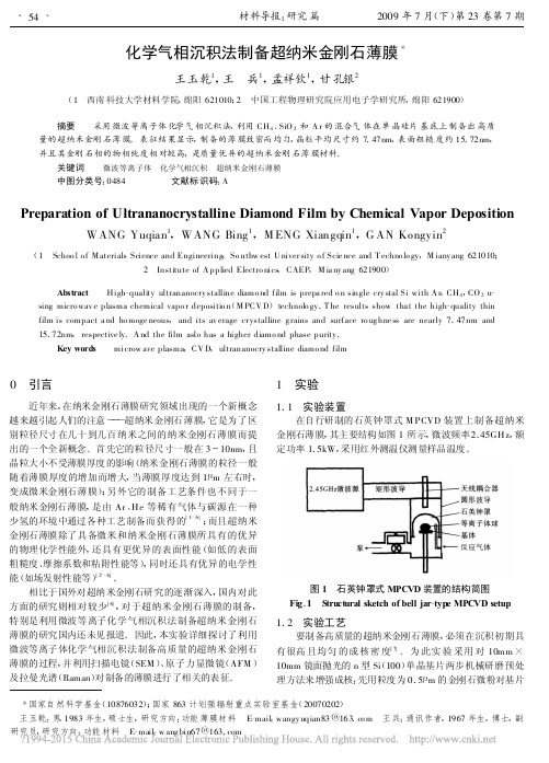
化学气相沉积法制备超纳米金刚石薄膜*王玉乾1,王 兵1,孟祥钦1,甘孔银2(1 西南科技大学材料学院,绵阳621010;2 中国工程物理研究院应用电子学研究所,绵阳621900)摘要 采用微波等离子体化学气相沉积法,利用CH 4、SiO 2和A r 的混合气体在单晶硅片基底上制备出高质量的超纳米金刚石薄膜。
表征结果显示,制备的薄膜致密而均匀,晶粒平均尺寸约7.47nm ,表面粗糙度约15.72nm ,并且其金刚石相的物相纯度相对较高,是质量优异的超纳米金刚石薄膜材料。
关键词 微波等离子体 化学气相沉积 超纳米金刚石薄膜中图分类号:0484 文献标识码:APreparation of Ultrananocrystalline Diamond Film by Chemical Vapor DepositionWANG Yuqian 1,WANG Bing 1,M ENG Xiangqin 1,G AN Kongyin2(1 Schoo l o f M aterials Science and Engineering ,So uthw est U niver sity o f Scie nce and T echno lo gy ,M iany ang 621010;2 Institute of A pplied Electro nics ,CAEP ,M ia ny ang 621900)Abstract High -quality ultrananocry stalline diamo nd film is prepa red o n single cry stal Si with A r ,CH 4,CO 2u -sing micro wav e plasma chemical vapo r depositio n (M PCV D )technolo gy .T he results show tha t the high -quality thin film is compact a nd ho moge neous ,and its av erage cr ystalline g rains and surface ro ug hne ss are nearly 7.47nm and 15.72nm ,respective ly .A nd the film aslo has a higher diamo nd phase purity .Key words microw ave plasma ,CV D ,ultrananocry stalline diamo nd film *国家自然科学基金(10876032);国家863计划强辐射重点实验室基金(20070202) 王玉乾:男,1983年生,硕士生,研究方向:功能薄膜材料 E -mail :wangy uqian83@163.co m 王兵:通讯作者,1967年生,博士,副研究员,研究方向:功能材料 E -mail :w ang bin67@0 引言近年来,在纳米金刚石薄膜研究领域出现的一个新概念越来越引起人们的注意———超纳米金刚石薄膜,它是为了区别粒径尺寸在几十到几百纳米之间的纳米金刚石薄膜而提出的一个全新概念。
Nanomaterials1.Introduction1、HistoryThe concept of a nanoscale technology begins with theboldly speculative 1959 article ‘There’s Plenty of Room atthe Bottom—An Invitation to Enter a New Field of Physics’by Nobel Prize winning theoretical physicist RichardFeynman. (We can arrange the atoms the way that wewant.)2、AFM-atomic force microscopeSTM-scanning tunneling microscopeCVD-chemical vapour deposition3、The properties of materials can be different at thenanoscale for two main reasons:First, nanomaterials have a relatively large surface area.Second, quantum effect can begin to dominate.2.New Materials: Nanomaterials1、Nanoscale in One DimensionThin films, layers and surfaces.2、Nanoscale in Two DimensionCarbon Nanotubes : CNTs are extended tubes ofrolled graphene sheets. There are two types of CNT:single-walled or mutil-walled .Inorganic NanotubesNanowires : nanowires are ultrafine(超细) wires or linear arrays of dots(线性点阵).Biopolymers(生物性聚合物)3、Nanoscale in Three DimensionNanoparticles(纳米颗粒):nanoparticle are often defined as particles of less than 100nm in diameter(直径).Fullerenes(carbon 60): In the mid-1980s a new class ofcarbon material was discovered called carbon 60(C60).Harry Kroto and Richard Smalley the experimental chemists who discovered C60 named it "buckminsterfullerene", inrecognition of the architect Buckminster Fuller, who waswell-known for building geodesic domes, and the termfullerenes was then given to any closed carbon cage.C60are spherical molecules about 1nm in diameter, comprising60 carbon atoms arranged as 20 hexagons(六边形) and12 pentagons(五边形): the configuration of a football in1990,a technique to produce larger quantities of C60 wasdeveloped by resistively heating graphite rods in a helium atmosphere. Several applications are envisaged forfullerenes, such as miniature ‘ball bearings’ to lubricatesurfaces, drug delivery vehicles and in electronic circuits.富勒烯(碳60):在20世纪80年代中期发现了一种新的碳材料,叫做碳60(C60)。

Characterizing the properties ofcarbon nanotubesCarbon nanotubes (CNTs) have been the subject of extensive research due to their unique structural, electronic, mechanical, and thermal properties. CNTs are cylindrical tubes of carbon atoms, having a diameter of a few nanometers and a length of several micrometers. The walls of CNTs are made of graphene sheets that are rolled up into cylinders, resulting in a seamless tube with a hollow core. The properties of CNTs depend on their diameter, length, chirality, and defects, which can be controlled during the synthesis process.One of the most important properties of CNTs is their high aspect ratio, which is the ratio of their length to diameter. CNTs can have aspect ratios of up to 100,000, which makes them the strongest known materials, with tensile strengths up to 63 GPa. The strength of CNTs comes from their sp2 hybridized carbon bonds, which make the tubes extremely stiff and resilient. CNTs are also highly flexible, and can bend and twist without breaking, enabling them to be used in a wide range of applications.Another important property of CNTs is their electrical conductivity. CNTs are excellent conductors of electricity, with an electrical conductivity of up to 1x10⁷ S/m, which is higher than that of copper. The conductivity of CNTs is dependent on their diameter and chirality, with smaller diameter tubes being more conductive than larger diameter tubes. The high conductivity of CNTs makes them a promising material for electronic and optoelectronic applications, such as transistors, sensors, and solar cells.CNTs also possess exceptional thermal conductivity, which is the ability to conduct heat. CNTs have an extremely high thermal conductivity of up to 3500 W/mK, which is higher than that of any other known material. The high thermal conductivity of CNTs makes them ideal for use in thermal management applications, such as heat sinks and nanocomposites.Furthermore, CNTs are highly hydrophobic, meaning that they repel water. This property makes them useful in applications where water resistance is required, such as in coatings and membranes. CNTs are also resistant to chemical corrosion and oxidation, which makes them highly durable and long-lasting.However, CNTs also have some limitations that need to be addressed. One of the major challenges is their toxicity. While CNTs have shown great promise in medical applications, such as drug delivery and cancer therapy, their potential toxicity to cells and tissues is a cause of concern. Studies have shown that CNTs can cause lung damage and inflammation in rodents, raising questions about their safety for human use. Therefore, it is important to thoroughly evaluate the toxicity of CNTs before using them in biomedical applications.In conclusion, CNTs are a remarkable material with unique and exceptional properties that make them suitable for a wide range of applications. Their high strength, electrical and thermal conductivity, hydrophobicity, and chemical stability make them a promising material in the fields of electronics, energy, and healthcare. However, their potential toxicity needs to be addressed before they can be widely used in biomedical applications. Understanding the properties of CNTs is essential for developing new applications that can exploit their exceptional properties while minimizing their drawbacks.。

国内图书分类号: TG 453国际图书分类号: 621.791工学博士学位论文超声楔形键合界面连接物理机理研究博士研究生:计红军导 师:王春青教授副 导 师:李明雨教授申请学位:工学博士学科、专业:材料加工工程所在单位:材料科学与工程学院答辩日期:2008年6月授予学位单位:哈尔滨工业大学Classified Index: TG 453U.D.C.: 621.791Dissertation for the Doctoral Degree in Engineering STUDY ON JOINING PHYSICAL MECHANISM OF ULTRASONIC WEDGE BOND INTERFACECandidate:Ji HongjunSupervisor:Prof. Wang ChunqingVice supervisor: Prof. Li MingyuAcademic Degree Applied for:Doctor of Engineering Specialty:Material Processing Engineering Affiliation: Department of Material Sci. & Eng. Date of Defence:June, 2008Degree-Conferring-Institution:Harbin Institute of Technology摘要摘要微电子、光电子系统中芯片级封装超声互连接头界面接合机制问题严重困扰着超声键合设备和技术的创新方向,也直接影响着元器件性能和可靠性。
因此,解析超声键合接头界面特征并厘清超声对接合过程所起到的本质作用对于指导封装互连设备升级、提高超声键合技术能力和改善元器件功能及寿命都具有重大意义。
本文针对常温超声楔形键合25μm Al-1wt.%Si引线与薄Au层Au/Ni/Cu、厚Au层Au/Ni/Cu、Cu三种焊盘之间形成的接头,原位测量了接头电阻;揭示了接头接合部连接物理过程和特征;考察了Au/Al和Al/Au 系统高温老化时接头界面演变特点;运用聚束离子束-透射电子显微镜(FIB-TEM)方法,在纳观尺度上给出了接头界面构成情况,在原子尺度上观察了界面接合特征;基于固相扩散反应原理,全面阐释了超声楔形键合接头界面弥散有纳米Au8Al3颗粒的固溶体形成过程中超声振动作用的物理实质。

磁性四氧化三铁纳米粒子的超声波辅助水热合成及表征*郭 英,李 酽,刘秀琳,才 华(中国民航大学理学院,天津300300)摘 要:以六水三氯化铁、四水二氯化铁和氨水为原料,在超声波辅助下,水热法制备了磁性四氧化三铁纳米粒子。
借助X射线衍射仪(XRD)、透射电镜(TE M)及振动样品磁强计(V S M)对产物四氧化三铁进行分析。
结果表明,当反应物三价铁离子与二价铁离子物质的量比为1.75,p H为13,控制水热合成温度在140~160 、水热处理时间在3~5h时,可制备出纯相的反尖晶石结构的四氧化三铁纳米微粒。
随着水热合成温度的升高和时间的延长,晶体发育更完整,平均粒径增大。
磁性测量表明,饱和磁化强度和矫顽力也随着四氧化三铁纳米微粒平均粒径的增大而增大,控制水热合成温度140~150 、水热处理时间4h能够制备出平均粒径小于20n m、具有超顺磁性的四氧化三铁纳米微粒。
关键词:纳米粒子;四氧化三铁;超声波处理;超顺磁性;水热合成中图分类号:TQ138.11 文献标识码:A 文章编号:1006-4990(2007)03-0021-04The ultrasonic wave assisted hydrot her m al synt hesis and charact erizationof nano-sized m agnetic Fe3O4particlesGuo Y i n g,LiY an,L i u X iuli n,C aiH ua(Colle ge of Sciences,C i vil A vi a tion Universit y of China,T ianjin300300,China)Abstrac t:The nano-sized m agnetic F e3O4parti c lesw as synthes i zed by hydro-t her m a lm ethod w it h the help of ultrason ic w ave by reacti ng FeC l3 6H2O w ith FeC l24H2O and NH3H2O.T he produced nano-sized m agnetic F e3O4partic les w as character ized by X-ray d iffraction(XRD),trans m issione lectron m icroscope(T E M)and v i bra ti ng sa m ple m agneto m eter(V S M).The exper i m enta l results sho w ed tha t F e3O4nano crysta l of m i s-spi ne l structure w ith high purity w as obta i ned at t he adopted n(F e3+)/n(F e2+)=1.75,p H=13,3~5h and140~160 .T he m ore i ntac t and l arger Fe3O4nanocrysta l was obta i ned w it h t he i ncreasi ng temperature and reacti on ti m e.T he V S M resu lts show ed t hat m agne tic saturati on m o m ent and coerciv i ty also w ere l a rger w it h the i ncre m en t o f t he ave rage g ra i n size o f Fe3O4nanocry sta.l The Fe3O4nanocrysta l w ith superpara m agnetic and av erage gra i n size under20n m cou l d be obta i ned,if contro l the hydrother m a l syn t hesis te m pe ra t ure at140~150 for4h.K ey word s:nano-pa rti c l es;Fe3O4;ultrasonic wave trea t m ent;s uperparam agnetic;hydro therma l synthesis目前,用于制备纳米磁性四氧化三铁的方法很多,如沉淀法、氧化还原法以及采用高温煅烧、火焰分解、激光热分解等干法合成,但是这些方法很难完全控制Fe2+的氧化,即Fe2+的氧化过程及比例,容易生成杂相;或使用的温度过高,使得晶粒生长过大,造成晶格缺陷的形成和杂质的引入。
纳米材料医学英语Nanotechnology in Medicine: Revolutionizing HealthcareThe field of medicine has witnessed remarkable advancements in recent decades, and one of the most promising areas of innovation is the use of nanotechnology. Nanotechnology, the science of manipulating matter at the atomic and molecular scale, has the potential to transform the way we approach healthcare, from diagnosis to treatment.The unique properties of nanomaterials, which are materials with dimensions in the nanometer range (1-100 nanometers), offer unprecedented opportunities in the medical field. These materials exhibit enhanced physical, chemical, and biological characteristics compared to their bulk counterparts, making them highly versatile and adaptable for various medical applications.One of the primary areas where nanotechnology is making a significant impact is in the field of drug delivery. Conventional drug delivery methods often face challenges such as limited bioavailability, undesirable side effects, and the inability to target specific tissues or cells. Nanomaterials, however, can be engineered to encapsulatedrugs, improving their solubility, stability, and targeted delivery. This can lead to more efficient and effective treatments, with reduced side effects and improved patient outcomes.For example, nanoparticles can be designed to carry and release drugs in a controlled manner, ensuring that the therapeutic agent reaches the intended site of action. These nanocarriers can be functionalized with targeting ligands, such as antibodies or peptides, which can recognize and bind to specific receptors on diseased cells, allowing for more precise and selective drug delivery. This targeted approach can enhance the efficacy of treatments while minimizing the exposure of healthy tissues to the drug, reducing the risk of adverse effects.Another exciting application of nanotechnology in medicine is the development of diagnostic tools and sensors. Nanomaterials can be engineered to interact with biological molecules, such as proteins, DNA, or cells, in highly sensitive and specific ways. This enables the creation of highly accurate and rapid diagnostic tests that can detect the presence of various biomarkers or pathogens at an early stage, allowing for timely interventions and improved patient outcomes.One example of this is the use of nanobiosensors, which can be designed to detect specific biomolecules or changes in the body's physiological conditions. These sensors can be integrated intowearable devices or implanted in the body, providing continuous monitoring and real-time data on the patient's health status. This can aid in the early detection of diseases, facilitate personalized treatment plans, and enable remote patient monitoring, improving the overall quality of healthcare delivery.Nanotechnology is also revolutionizing the field of regenerative medicine, where the goal is to repair, replace, or regenerate damaged or diseased tissues and organs. Nanomaterials can be used to create scaffolds that mimic the extracellular matrix, providing a suitable environment for cell growth and tissue regeneration. These scaffolds can be loaded with growth factors, stem cells, or other therapeutic agents to enhance the body's natural healing processes.Furthermore, nanomaterials can be designed to interact with the immune system in a way that modulates the body's response to injury or disease. For instance, nanoparticles can be engineered to suppress the inflammatory response or to stimulate the immune system to fight against specific pathogens or cancer cells, opening up new avenues for the treatment of various diseases.The potential of nanotechnology in medicine is not limited to these examples. Researchers are also exploring the use of nanomaterials in medical imaging, tissue engineering, and the development of smart materials that can adapt to changing physiological conditions. Theseadvancements hold the promise of improving the accuracy of diagnoses, enhancing the efficacy of treatments, and ultimately, improving the overall quality of life for patients.However, the integration of nanotechnology into healthcare is not without its challenges. Ensuring the safety and biocompatibility of nanomaterials is a critical concern, as their small size and unique properties may interact with biological systems in unpredictable ways. Rigorous testing and regulatory oversight are necessary to ensure the safe and ethical development and application of nanomedicine.Despite these challenges, the future of nanotechnology in medicine is bright. As research continues to advance, we can expect to see even more remarkable breakthroughs that will transform the way we approach healthcare, leading to personalized, precise, and more effective treatments. The integration of nanotechnology into the medical field holds the potential to revolutionize the way we diagnose, treat, and manage a wide range of health conditions, ultimately improving the overall well-being of individuals and communities around the world.。


中国病原生物学杂志2020年12月第15卷第12期•1370•Journal of Pathogen Biology Dec.2020,Vol.15.No.121)01:10.13350/j.cjpb.201202•论著•一株野鸟源H16N3亚型禽流感病毒的遗传进化分析与感染能力评估*孙雷云李元果张醒海….赵梦琳;.胡鑫宇;.王铁成‘,孙伟洋',冯娜:赵永坤杨松涛夏成柱「,孟德荣‘.高玉伟心…(1•占林农业大学动物科学技术学院,吉林长春130118;2.军事医学研究院军事兽医研究所;3.吉林大学;4.沧州师范学院〉目的了解H16N3亚型禽流感病毒的遗传进化特征及其化物学特性,为野鸟源禽流感病毒预警提供科学依据。
方法采集途径我国中东部地区重要候鸟栖息地的野鸟粪便样品.经处理后接种SPF鸡胚.获得具有血液凝集特性病原体.经全底因测序确定病毒亚型。
选取H16亚型流感病毒构建系统发育树并进行分子特性分析。
检测病毒受体结合特性.并进行小鼠和家禽感染试验.评价该病毒对哺乳动物和家禽的致病性。
结果分离到1株病原体(CZ-638),经全基因组测疗;及电镜观察.确定为H16N3W:型禽流感病毒。
在系统发育树种,该带株位于欧亚谱系分支。
氨基酸位点分析显示.HA蛋白裂解位点为INERl GI.F.符合低致病性禽流感病毒分子特征.受体结合域的228位点由G (It氨酸)突变为S"纟訊酸)。
该病毒株能够凝集绵羊红细胞、正常鸡红细胞及仅有SA«2,6受体的鸡红细胞.表明该毒株具有双受体结合能力。
动物感染试验显示.该毒株对小鼠、1周龄雏鸡、亚成体家鸭均不具有感染力。
结论分离的H16N3毒株为欧亚谱系.对小鼠和家禽无致病性。
该毒株存在结合人1:呼吸道流感病毒受体的能力.但尚未获得感染家禽和哺乳动物的能力•应持续监测.追踪病毒进化待征°关键词】禽流感病馭H16N3;遗传进化;致病性;受体结合待性;感染能力中图分类号】S852.65【文献标识码】【文章编号】1673-5234(2020)12-1370-07[Journal of Pathogen Biology.2020Dec;15(12):1370—1376.]A genetic evolutionary analysis and an evaluation of the infectivity of avian influenza H16N3isolated fromwild birds in ChinaSUN Lei-yun1'・LI Yuan-guo,ZHANG Xing-hai2',ZHAO Meng-lin・HU Xin—yu,WANG Tie-cheng J,SUN Wei-yang・FENG Na2・ZHAO Yong-kun~・YANG Song-tao2•XIA Xian-zhu2,MENG De~R o n g1,GAO Y u-wei~(1.College of Alli mal Science and Technology・Jilin Agricultural University»Changchun9China130118; 2.Institute of Military Veterinary Medicine.Academy of Military Medical Science; 3.J ilin University; 4.Can^zhou Normal University)Objectives To ascertain the genetic evolutionary characteristics and biological characteristics of the H16N3 subtype of the avian influenza virus in order to provide a scientific basis for early warning of avian influenza virus from wild birds.Methods Fecal samples from wild birds in major migratory bird habitats in central and eastern China were collected and inoculated into SPF chicken embryos after treatment to obtain pathogens with blood agglutination characteristics.and the virus subtypes were determined using whole gene sequencing.A phylogenetic tree was constructed for the H16subtype of the influenza virus.and the subtypes were characterized molecularly.The receptor binding characteristics of the virus were determined and infection tests were conducted in mice and poultry to evaluate the pathogenicity of the virus to mammals and poultry.Results One strain of pathogen(CZ-638)was isolated and identified as avian influenza virus subtype H16N3according to whole genome sequencing and electron microscopy.In the phylogenetic tree・the strain was located in the Eurasian lineage branch.An analysis of amino acid sites indicated that an HA protein cleavage site was INER J GLF.which was in line with the molecular characteristics of the low pathogenic avian influenza virus.Amino acid 228of the receptor binding domain mutated from G(glycine)to S(serine).This strain agglutinated sheep red blood cells・normal chicken red blood cells,and chicken red blood cells with the SAa2,6receptor alone・indicating that this vi-【基金项目】【通讯作者】【作者简介】国家科技重大专项(No.2O2OZX1OOO1-O16-OO3);国家自然科学基金项目(No.31970502)。
Nanocrystalline materials and coatingsS.C.Tjong *,Haydn ChenDepartment of Physics and Materials Science,City University of Hong Kong,Tat Chee Avenue,Kowloon,Hong Kong Available online 10August 2004AbstractIn recent years,near-nano (submicron)and nanostructured materials have attracted increasingly more attention from the materials community.Nanocrystalline materials are characterized by a microstructural length or grain size of up to about 100nm.Materials having grain size of $0.1to 0.3m m are classified as submicron materials.Nanocrystalline materials exhibit various shapes or forms,and possess unique chemical,physical or mechanical properties.When the grain size is below a critical value ($10–20nm),more than 50vol.%of atoms is associated with grain boundaries or interfacial boundaries.In this respect,dislocation pile-ups cannot form,and the Hall–Petch relationship for conventional coarse-grained materials is no longer valid.Therefore,grain boundaries play a major role in the deformation of nanocrystalline materials.Nanocrystalline materials exhibit creep and super plasticity at lower temperatures than conventional micro-grained counterparts.Similarly,plastic deformation of nanocrystalline coatings is considered to be associated with grain boundary sliding assisted by grain boundary diffusion or rotation.In this review paper,current developments in fabrication,microstructure,physical and mechanical properties of nanocrystal-line materials and coatings will be addressed.Particular attention is paid to the properties of transition metal nitride nanocrystalline films formed by ion beam assisted deposition process.#2004Elsevier B.V .All rights reserved.Keywords:Nanostructure;Synthesis;Mechanical properties;Grain size;Processing-structure–property;Nanocrystalline coatings;Ion beam1.IntroductionNanomaterials are experiencing a rapid development in recent years due to their existing and/or potential applications in a wide variety of technological areas such as electronics,catalysis,ceramics,magnetic data storage,structural components etc.To meet the technological demands in these areas,the size of the materials should be reduced to the nanometer scale.For example,the miniaturization of functional electronic devices demands the placement or assembly of nanometer scale components into well-defined structures.As the size reduces into the nanometer range,the materials exhibit peculiar and interesting mechanical and physical properties,e.g.increased mechanical strength,enhanced diffusivity,higher specific heat and electrical resistivity compared to conventional coarse grained counterparts [1].Nanomaterials can be classified into nanocrystalline materials and nanoparticles.The former are polycrystalline bulk materials with grain sizes in the nanometer range (less than 100nm),while the latter refers to ultrafine dispersive particles with diameters below 100nm.Nanoparticles are generally considered as the building blocks of bulk nanocrystalline materials.Research in nanomaterials is a multidisciplinary effort that involves interaction between researchers in the field of physics,chemistry,mechanics and materials science,or even biology and medicine.It has been stimulated bythe Materials Science and Engineering R 45(2004)1–88*Corresponding author.Tel.:+852********;fax:+852********.E-mail address:aptjong@.hk (S.C.Tjong).0927-796X/$–see front matter #2004Elsevier B.V .All rights reserved.doi:10.1016/j.mser.2004.07.0012S.C.Tjong,H.Chen/Materials Science and Engineering R45(2004)1–88 interest for basic scientific investigations and their technological applications.Nanomaterials and mostof the applications derived from them are still in an early stage of technical development.There areseveral issues that remain to be addressed before nanomaterials will become potentially useful forindustrial sectors.These issues include synthesis of high purity materials with large yield economicallyand environmentally,characterization of new structures and properties of nanophase materials,fabrication of dense products from nanoparticles with full density and less contamination,and retentionof the ultrafine grain size in service in order to preserve the mechanical properties associated with thenanometer scale.Novel fabrication technology of nanoparticles is versatile and includes a wide range of vapor, liquid and solid state processing routes.Available techniques for the synthesis of nanoparticles viavapor routes range from physical vapor deposition and chemical vapor deposition to aerosol spraying.The liquid route involves sol–gel and wet chemical methods.The solid state route preparation takesplace via mechanical milling and mechanochemical synthesis.Each method has its own advantagesand shortcomings.Among these,mechanical milling and spray conversion processing are commonlyused to produce large quantities of nanopowders.Nanoparticles synthesized from several routes may have different internal structures that would affect the properties of materials consolidated from them.Processing nanoparticles into fully dense,bulk products or coatings which retain the nanometer scale grain size is rather difficult to achieve inpractice.Due to their high specific surface areas,nanoparticles exhibit a high reactivity and strongtendency towards agglomeration.Moreover,rapid grain growth is likely to occur during processingat high temperatures.As unique properties of nanocrystaline materials derived from theirfinegrain size,it is of crucial importance to retain the microstructure at a nanometer scale duringconsolidation to form bulk materials.It is also noticed that pores are generated in bulk nanocrystal-line materials consolidated from nanoparticles prepared by inert-gas condensation.Such nano-pores can lead to a decrease in Young’s modulus of consolidated nanocrystalline materials[2].Electrodeposited samples are believed to be free from porosity,but they contain certain impuritiesand texture that may degrade their mechanical performances.Therefore,controlling these proper-ties during synthesis and subsequent consolidation procedures are the largest challenges facingresearchers.The unique properties of nanocrystalline materials are derived from their large number of grain boundaries compared to coarse-grained polycrystalline counterpartes.In nanocrystalline solids,alarge fraction of atoms(up to49%)are boundary atoms.Thus the interface structure plays animportant role in determining the physical and mechanical properties of nanocrystalline materials.Huang et al.[3]reported that nanocrystalline copper has a much higher resistivity and a largertemperature dependence of the resistivity than bulk copper.They attributed this effect to the grain-boundary enhanced scattering of electrons.Nanocrystalline metals have been found to exhibit creepand superplasticity with high strain rates at lower temperatures than their micro-grained counterparts.High strain-rate superplasticity at lower temperatures is of practical interest because it can offer anefficiently near-net-shape forming technique to industrial sectors.Despite recent advances in thedevelopment of nanocrystalline materials,much work remains to be done to achieve a basicunderstanding of their deformation and fracture behavior.Nanocrystalline metals generally exhibitsignificantly higher yield strength and reduced tensile elongation relative to their microcrystallinecounterparts.The hardness and yield strength tend to increase with decreasing grain size down to acritical value(ca.20nm).When the grain size is below20nm,strength appears to decrease withfurther grain refinement.At this stage,dislocation sources inside the grains can hardly exists.Thisimplies that dislocation pile-ups cannot form and the Hall–Petch relationship for conventional coarser-grained materials is no longer valid.Instead,inverse Hall–Petch effect,i.e.softening is obtained whenS.C.Tjong,H.Chen/Materials Science and Engineering R45(2004)1–883 the grain size is reduced.The softening behavior of nanocrystalline materials is a subject of considerable debate.Several mechanisms have been proposed to explain the anomalous deformationbehavior of nanocrystalline materials with the grain size below the critical value.These include grain-boundary sliding,grain-boundary diffusion,the triple junction effect,presence of nanopores and impurities,etc.Therefore,comprehensive understanding of the processing-structure–property rela-tionships is essential in the development of novel nanomaterials with unique properties for structural engineering applications.As dense nanocrystalline materials with grain size smaller than20nm aredifficult to acquire,molecular dynamics(MD)modeling has been used to simulate the interfacialstructure and mechanical deformation mechanism of such puter simulations play acritical role in advancing our understanding of atomic level and deformation structures that are not accessible by experimental routes.Nanocrystalline coatings with grain sizes in the nanometer range are also known to exhibitsuperior hardness and strength.The search for nanostructured coatings is driven by the improvement incoating technologies and the availability of various kinds of synthesized nanopowders.Such nanopowders can be used as feedstock materials for thermal spray processes;these include plasmaspraying and high-velocity oxygen fuel(HVOF)spraying.Thermal spraying involves particle melting,rapid cooling and consolidation in a single-step operation.Thermal-sprayed nanocrystalline coatingswith moderate hardness are found to possess better wear performances than their counterparts fabricated from microcrystalline powders.HVOF is particularly suited to deposit dense nanocrystal-line ceramic coatings as opposed to plasma spraying because of its lower spraying temperature.Today,HVOF allows tailoring nanocrystalline coatings with low porosity,higher bond strength and increasedwear properties.In the past decade,favorable applications have been found for hard and wear-resistant ceramiccoatings in industrial sectors.Transition metal nitride coatings are of particular interest due to theirhigh hardness,thermal stability,attractive appearance and chemical inertness.Conventional nitridecoatings have been prepared by physical and chemical vapor deposition.Nanocrystalline coatings of transition metal nitrides can be deposited on substrate materials by means of ion beam assisted deposition.The process is based on simultaneous ion bombardment of the growing physical vapor depositedfilm using an independent ion source.This technique permits the deposition of nanocrystal-line nitridefilms at lower temperatures with better coating-substrate adhesion.The improvement of tool materials coated with transition metal nitrides has led to interest in developing superhard coatings for wear protection under complex loads and aggressive environments.Optimal microstructural design and materials selection permit chemical,physical and mechanical characteristics can be tailored for specific applications.Superhard coatings having hardness valuesabove40GPa are obtainable in multilayer structures with the period of the superlattice(bilayer thickness)within the nanometer regime.The enhancement of superhardness is attributed to a difference in shear modulus between two layer materials and to the presence of sharp interfacesbetween the layers.In another approach,nanocomposite coatings with superhardness!40GPa can beprepared by dispersing the transition metal nitride nanoparticles in an amorphous covalent nitridematrix(1nm).In terms of MD computer simulations,grain-boundary accommodation mechanismssuch as grain-boundary sliding and diffusion are considered to be the main factors causing the softening of nanocrystalline materials with grain sizes below10nm.Blocking of grain-boundarysliding of nanocrystalline grains embedded in a thin amorphous matrix is believed to be responsible for superhardness of the nanocomposite coatings.Because of the multidsiplinary nature of nanomaterials,it would be difficult to cover all areas of interest.In this review article,we address and focus the discussions on the following subjects,namely nanoparticles,nanocrystalline materials and coatings.2.NanostructureOne of the most critical characteristics of nanoparticles is their very high surface-to-volume ratio,rge fractions of surface atoms.Thus,large fractions of surface atoms together with ultra-fine sizeand shape effects make nanoparticles exhibit distinctly different properties from the bulk.Theevolution of nanoparticles from a vapor or liquid phase involves three fundamental steps:nucleation,coalescence and growth.When the concentration of building blocks(atoms or ions)of a solid becomessufficiently high,they aggregate into small clusters through homogeneous nucleation.With con-tinuous supply of the building blocks,these clusters tend to coalesce and grow to form a larger clusterassembly.Thus,the nanoparticles are often built-up from a full-shell cluster of atoms having cubic orhexagonal closed-packed structure.Such a structure can be constructed from a central atomsurrounded by afirst shell of12,a second of42,a third of92atoms,etc.The number of atomsin nth shell is10n2+2(Table1).The coordination number of surface atoms is9or smaller,anddifferent from their bulk counterparts,i.e.12.With decreasing size of the nanoparticles,the percentage 4S.C.Tjong,H.Chen/Materials Science and Engineering R45(2004)1–88 Table1The relation between the total number of atoms in full shell clusters and the percentage of surface atoms(reprinted from[5]with permission from John Wiley&Sons)Full shell clusters Total number of atoms Surface atoms(%)Oneshell1392Twoshells5576Threeshells14763Fourshells30952Fiveshells56145Sevenshells141535of surface atoms increases.The critical role of the size of nanoparticles in physical properties has been demonstrated experimentally in melting temperature.For example,Fig.1shows the typical relation between the particle size and melting point of gold particles,calculated by the method of Reifenberger and coworkers [4].Apparently,the melting temperature of gold reduces dramatically from 1063to $3008C for nanoparticles with diameters of smaller than 5nm [5].In general,the geometrical shape is determined by the composition and properties of the synthesized material,or by the formation mechanism of the condensed nanoparticles.For most transition metal nanoparticles,distinct structures can occur that are not characteristic of the bulk crystal structure.These structures are:cubooctahedron,icosahedron and decahedron (Fig.2).The first two structures are commonly observed in clusters of gold,Cr and other metals [6,7].Clusters with a small number of atoms (<150–200)crystallize in the form of icosahedra.The structure becomes unstable for a large number of atoms and transforms to cubooctahedra,which is just a patch of the face-centered-cubic (fcc)lattice [8,9].A typical example is the change from icosahedral to face-centered cubic structure in gold particles [9].The particle shapes are closely related to the crystal-lographic surfaces that enclose the particles.The {111},{100}and possibly {110}surfaces of fcc metal particles,are different not only in the surface atom densities but also the electronic structure,bonding and chemical reactivities [10].Controlling the size,shape and structure of metal nanoparticles is technologically important because of strong correlation between these parameters and optical,electrical and catalytic properties [11].Many metals can now be processed into monodisperse nanoparticles with controlled composition and structure.They can be produced in large quantities through solution phase methods.Other morphologies with less stable facets have been achieved by adding chemical capping reagents to the synthetic systems [11–13].For example,the shapes and sizes of Pt (fcc)nanoparticles such as cubic,tetrahedral and octahedral can be controlled by changes in the ratio of the concentration of the caping polymer material (sodium polyacrylate)to the concentration of the Pt ions used in the reductive synthesis of colloidal particles in solution at room temperature.The presence of the polymer in the solution of colloids is believed to have two functions.First,it stops the growth of the particles at a small size distribution.Second,it prevents individual colloidal particles from coalescing with each other [13].The fcc nanocystals normally have {111}twins.The fc metals tend to nucleate and grow into twinned and multiply twinned particles (MTP)with their surfaces bounded by the lowest energy {111}facets [14].Twining is the result of two subgrains sharing a common crystallographic plane.In this case,the structure of one subgrain is the mirror re flection of the S.C.Tjong,H.Chen /Materials Science and Engineering R 45(2004)1–885Fig.1.Relation between the size of gold particles and their melting point (reprinted from [5]with permission from John Wiley &Sons).other by the twin plane.The two most typical examples of MTP are decahedron and icosahedron.As demonstrated by Wang [10],the shape of an fcc crystal is mainly determined by the ratio (R )between the growth rates in the (100)direction to that of the (111).Octahedra and tetrahedral bounded by the most stable {111}planes will be formed when R =1.73.Perfect cubes bounded by the less stable {100}planes will result if R is reduced to 0.58.The particles with 0.87<R <1.73have the {100}and {111}facets,which are referred to truncated octahedron (Fig.2).High resolution transmission electron microscopy (HRTEM)is generally known as a powerful tool to observe the morphology and structure of nanoparticles This is due to its atomic resolution capabilities to observe spatially resolved chemistry at nanoscale [15–19].Wang et al.employed HRTEM to directly image atomic scale structures of the surfaces of platinum particles with cubic-,octahedral-and tetrahedral-like shapes prepared from a shape-controlling technique [10,19].Fig.3a and b shows typical HRTEM images of cubic Pt nanocrystals oriented along [001]and [110]directions,respectively.From Fig.3a,the particle is bounded by {100}facets and there is no defect in the bulk of the particle.The distance between the adjacent lattice fringes is the interplanar distance of Pt (200),which is 0.196nm.The particle surface has some steps and ledges,particularly near the corner of the cube.Fig.4a shows the HRTEM image of a truncated octahedral Pt nanoparticle oriented along [110].The HRTEM image of the octahedral-like Pt nanocrystals oriented along [110]and [001],respectively are shown in Fig.4b[19].An octahedron has eight {111}facets,four of them are edge-on when viewed along [110].If the particle is a truncated octahedron,six {100}facets are created by cutting the corners of octahedron,two of which are edge-on while viewing along [110].The properties of bulk nanocrystalline materials are known to be controlled by their ultra-fined grain sizes and interface boundaries.Small grain size in nanocrystalline materials produces large interfacial regions per unit volume as expected.The atomic arrangements or structures of the grain boundaries have been intensively investigated in the past decades.However,controversial results in the grain boundary structures are obtained.Some researchers suggested that the grain boundary 6S.C.Tjong,H.Chen /Materials Science and Engineering R 45(2004)1–88Fig.2.(a)Geometrical shapes of cubooctahedral nanocrystals as a function of the ratio,R ,of the growth rate along the (100)to that of the (111).(b)Evolution in shapes of a series of (111)based nanoparticles as the ratio {111}to {100}increases.The beginning particle is bounded by three {100}facets and a (111)base,while the final one is a {111}bounded tetrahedron.(c)Geometrical shapes of multiply twinned decahedral and icosahedral particles (reprinted from [10]with permission from American Chemical Society).structure is similar to that in coarser-grained materials [20–30],while others proposed a frozen ‘gaslike ’structure of the grain boundaries in which the atomic arrangement at interfacial region lacks short-or long-range order [31–33].In nanocrystalline solids a large fraction of atoms are located near grain boundaries.Assuming a grain boundary thickness of 1nm,materials with an average grain S.C.Tjong,H.Chen /Materials Science and Engineering R 45(2004)1–887Fig.3.HRTEM micrographs of cubic Pt nanoparticles oriented along (a)[001]and (b)[110],showing surface steps or ledges (reprinted from [19]with permission fromElsevier).Fig.4.HRTEM image of Pt nanoparticles with a (a)truncated octahedral shape and oriented along [110];(b)with an octahedral shape and oriented along [110]and [001](reprinted from [19]with permission from Elsevier).diameter of 5nm possesses $49%grain boundary atoms (Fig.5).Earlier studies of the interfacial structures of nanocrystalline Pd with HRTEM [6]and X-ray diffraction techniques [27,28]as well as nanocrystalline Cu with EXAFS [27]indicated that the atomic displacements in the grain boundaries were very small,and the structures were similar to those in coarser-grained materials.Fig.6is the HRTEM image of the interfacial grain boundaries in nanocrystalline Pd showing the morphology of flat facets interspersed with steps [22].On the basis of X-ray diffraction results,Gleiter and coworkers 8S.C.Tjong,H.Chen /Materials Science and Engineering R 45(2004)1–88Fig.5.Range of percentage of atoms in grain boundaries of a nanocrystalline solid as function of grain diameter,assuming that the average grain boundary thickness ranges from 0.5to 1.0nm (reprinted from [20]with permission from Annual ReviewInc.)Fig.6.HRTEM image of a region of nanocrystalline palladium containing a number of grains (reprinted from [22]with permission from Elsevier).reported that the grain boundaries of nanocrystalline pure Fe exhibiting a strongly reduced local atomic short-range order.This implies that the atomic structure in grain boundaries of nanocrystalline materials is different from that in conventional coarse-grained polycrystals [30].Gleiter proposed a ‘hard-sphere ’two-dimensional model for a nanocrystalline solid as shown in Fig.7[28].Two different structures of atoms are illustrated in the diagram:crystal atoms with neighbor con figuration corresponding to the lattice and boundary atoms with a wide variety of interatomic spacings.In the boundary regions,the coordination between nearest neighbor atoms deviates or reduces from the one in the crystallites,implying the occurrence of atomic disorder at these regions [28,34].Further,Gleiter demonstrated that several factors like size effects,changes of the atomic structure,and alloying elements could affect the properties of nanocrystalline materials.The possible effect of alloying elements on the atomic arrangements of nanocrystalline alloys is illustrated in Figs.8and 9.Solute atoms with little solubility in the lattice of the crystallites often segregate to the boundary regions [1].In certain cases,the formation of a Ag-Fe alloy solid solution is favored even though the constituents are immiscible in the crystalline and molten state (e.g.Fe and Ag)[32].S.C.Tjong,H.Chen /Materials Science and Engineering R 45(2004)1–889Fig.7.Two-dimensional model of a nanocrystaline solid.The atoms in the center of the crystals are indicated in black.The ones in the boundary regions are represented as open circles (reprinted from [1]with permission fromElsevier).Fig.8.Schematic model of the structure of the nanocrystalline Cu –Bi and W –Ga alloys.The open circles represent the Cu or W atoms,respectively,forming the nanometer-sized crystals.The black circles are the Bi or Ga atoms,respectively,incorporated in the boundaries of alloys (reprinted from [1]with permission from Elsevier).Nanocrystalline materials can be classi fied into several groups according to their dimensionality:zero-dimensional atom clusters,one-dimensional modulated multilayers,two-dimensional ultra-fine-grained overlayers,and three-dimensional nanocrystalline structures (Fig.10)[35].The nanocrystal-line materials may contain crystalline,quasi-crystalline and amorphous (nanoglasses)phases.They can be metals,intermetallics,ceramics and composites.Gleiter [36,37]classi fied nanocrystalline solids into twelve groups according to the shape (dimensionality)and chemical composition of their constituent structural elements (Fig.11).According to the shape of crystallites,three categories of nanocrystalline materials may be distinguished:layer-shaped crystallites,rod-shaped crystallites (with layer thickness or rod diameters on the order of a few nanometers)and nanocrystalline materials composed of equiaxed nanometer-sized crystallites.Depending on the chemical composition of the crystallites,the three categories of nanocrystalline materials may be grouped into four families.In the first family,all crystallites and interfacial regions have the same chemical composition.The second 10S.C.Tjong,H.Chen /Materials Science and Engineering R 45(2004)1–88Fig.9.Schematic model of nanocrystalline Ag –Fe alloys that consist of a mixture of nanometer-sized Ag and Fe crystals (represented by open and full circles,respectively).In the interfacial regions between Ag and Fe crystals,solid solution of Fe atoms in Ag crystallites,and Ag atoms in the Fe crystallites are formed although both component are immiscible in the liquid as well in the molten stat (reprinted from [32]with permission fromElsevier).Fig.10.Schematic of the four types of nanocrystalline materials according to Siegel (reprinted from [35]with permission from Elsevier).family consists of crystallites with different chemical compositions.If the composition variation occurs primarily between crystallite and the interfacial regions,the third family is obtained.In this case,one type of atoms tends to segregate preferentially to the interfacial regions (see also Fig.8).The fourth family is formed by nanometer-sized crystallites (layers,rods,equiaxed crystallites)dispersed in a matrix of different chemical composition.Precipitation alloy,e.g.dispersion of Ni 3Al in Ni matrix is a typical example of this family.Grain growth is a crucial aspect of the thermal stability of nanocrystaline solids.Nanocrystalline materials are thermodynamically unstable due to the presence of a large fraction of interface boundaries.There is a strong tendency for nanocrystalline materials to convert to conventional coarser grained materials with fewer interfaces.Therefore,stabilization of the nanocrystalline grain structure is of critical importance for retaining their unique structures and properties.For conventional polycrystalline materials,the driving force for grain growth results from the the reductions of free energy of the system by decreasing the total grain boundary energy.The kinetic equation of grain growth can be described by the following equation:d 2Àd 20¼kt (E1)where d is the average grain size after annealing,d 0the initial grain size,t the annealing time,and k a material constant.However,the parabolic grain growth as expressed in Eq.(E1)is rarely observed except for high purity metals at high homologous temperatures.In most cases,the variation of grainFig.11.Classi fication schema for nanocrystalline materials according to their chemical composition and the dimensionality (shape)of the crystallites (structural elements)forming the materials (reprinted from [37]with permission from Elsevier).。

Trans. Nonferrous Met. Soc. China 28(2018) 1763−1773Osteogenic composite nanocoating based on nanohydroxyapatite,strontium ranelate and polycaprolactone for titanium implantsMurat Taner VURAT1,2, Ayşe Eser E LÇIN1, Yaşar Murat E LÇIN1,21. Tissue Engineering, Biomaterials and Nanobiotechnology Laboratory,Faculty of Science and Stem Cell Institute, Ankara University, Ankara 06100, Turkey;2. Biovalda Health Technologies, Inc., Ankara 06830, TurkeyReceived 16 October 2017; accepted 3 April 2018Abstract: Titanium and its alloys are commonly used as dental and bone implant materials. Biomimetic coating of titanium surfaces could improve their osteoinductive properties. In this work, we have developed a novel osteogenic composite nanocoating for titanium surfaces, which provides a natural environment for facilitating adhesion, proliferation, and osteogenic differentiation of bone marrow mesenchymal stem cells (MSCs). Electrospinning was used to produce composite nanofiber coatings based on polycaprolactone (PCL), nano-hydroxyapatite (nHAp) and strontium ranelate (SrRan). Thus, four types of coatings, i.e., PCL, PCL/nHAp, PCL/SrRan, and PCL/nHAp/SrRan, were applied on titanium surfaces. To assess chemical, morphological and biological properties of the developed coatings, EDS, FTIR, XRD, XRF, SEM, AFM, in-vitro cytotoxicity and in-vitro hemocompatibility analyses were performed. Our findings have revealed that the composite nanocoatings were both cytocompatible and hemocompatible; thus PCL/HAp/SrRan composite nanofiber coating led to the highest cell viability. Osteogenic culture of MSCs on the nanocoatings led to the osteogenic differentiation of stem cells, confirmed by alkaline phosphatase activity and mineralization measurements. The findings support the notion that the proposed composite nanocoatings have the potential to promote new bone formation and enhance bone-implant integration.Key words: osteogenic nanocoating; composite nanofiber; titanium implant; nanohydroxyapatite; strontium ranelate; polycaprolactone; electrospinning; mesenchymal stem cells1 IntroductionBone is a highly vascularized and dynamic organthat has a number of functions such as protection,mineral storage and source of hematopoietic andmesenchymal stem cells (MSCs). Chemically, bonetissue consists of 1% sodium chloride, 1%−2%magnesium phosphate, 1%−2% calcium fluoride, 7%calcium carbonate and 58% calcium phosphate. Bonetissue, known as osseous, is made of cancellous andcortical bone. Living bone is mainly composed ofcollagen (70%), bone mineral and water. In addition,organic materials such as polysaccharides, proteins andlipids are present in less amounts [1,2]. Bone healing is acomplex process with three distinct overlapping steps:early inflammatory stage, repair stage and the lateremodeling stage [3,4]. Initially, within few days a localhematoma develops at the fracture site. Cellularinflammatory elements such as lymphocytes, monocytes,macrophages, and polymorphonuclear cells infiltrate thehematoma to secrete cytokines and growth factors. In therepair stage, fibroblasts facilitate vascular ingrowth andthere is high osteoblast activity. At this stage, the newlyformed bone needs to be remodeled. The last stage ofbone tissue healing is the remodeling stage where bone isrestored to its healthy condition [5,6].The coating of orthopaedic implants or bone fixatorsurfaces aims to improve the implant-bone integration,restore tissue functions and accelerate bone healing. Anideal implant surface coating should mimic the bonetissue by being biocompatible, osteoinductive, andmechanically stable [6].Due to their high corrosion resistance, uniquemechanical properties, osseointegration capacity andhigh biocompatibility with the tissue, titanium and itsalloys are widely preferred in the dental and orthopaedicimplant applications [7,8]. Owing to their lack of directCorresponding author:Yaşar Murat E LÇIN; E-mail: elcinmurat@; elcin@.trDOI:10.1016/S1003-6326(18)64820-4Murat Taner VURA T, et al/Trans. Nonferrous Met. Soc. China 28(2018) 1763−1773 1764chemical bonding capacity with the tissue, titanium alloys are bioinert materials. Some surface modifications are applied to titanium alloys for providing better implant integration. Metals and their alloys are coated with various biocompatible materials for biomedical applications. Some coating techniques, such as sol−gel, thermal spraying, sputtering, dipping and immersion coating, and electrophoretic deposition, were used in previous works [9]. Besides, the electrospininng process is used in many biomedical applications. Electrospinning is useful for manufacturing non-woven polymer fibers with diameters in the range of submicrometers to nanometers [10−12].Polycaprolactone (PCL) is a prominent polymer which has been widely used in biomedical applications, due to its non-toxicity, good mechanical properties, slow degradation rate, low-cost and high biocompatibility. PCL has high thermal stability with a melting temperature of 58−60 °C, and a glass transition temperature of −60 °C. Also, PCL has already been approved for use as a biomaterial for a number of applications by the FDA. In addition to these advantages, electrospun PCL nanofibers could mimic the extracellular matrix (ECM) of many tissues [13−17].Chemical composition and physical structure of hydroxyapatite (HAp) are very similar to those of the natural bone, hence, HAp has been used in hard tissue engineering studies alone or as a biomaterial component [18−20]. Additionally, HAp has been used as a coating material for implant applications owing to its good biocompatibility, osteoconductivity and bioactivity [21,22]. HAp displays high binding capacity to bone and enhances bone tissue formation.Strontium is an essential trace element with bioactive properties related to the skeletal metabolism. Recent in-vitro and in-vivo studies have reported the unique beneficial properties of strontium ions on bone healing [23−25]. Supporting this notion, recent work has shown that strontium induces MSC differentiation and pre-osteoblast proliferation, while also contributing to bone homeostasis by inhibiting osteoclast activity [26]. More previous researches elucidated the mechanism of strontium on the differentiation of osteoprogenitor cells into bone-forming osteoblasts through regulation of Wnt/β-catenin signalling pathway and expression of membrane-bound calcium sensing receptor (CaSR) [27,28]. Further evidence supports the role of strontium in bone anabolism, i.e., inducing the decrease in osteoclastogenesis, hence significantly lowering resorption activity of mature osteoclasts [29,30]. Also, chemical and physical structures of strontium are similar to those of calcium and about 98% of the total body strontium content is localized in the bone tissue [31].Strontium ranelate (SrRan), a strontium chelating agent, is widely used for reducing bone fractures in high risk patients and for the treatment of osteoporosis, especially in postmenopausal women. In-vitro experiments have indicated a unique positive effect of SrRan on the bone tissue [32]. Also, SrRan can decrease osteoclast differentiation and promote osteoblast proliferation and activity. It also regulates osteoclast activity by editing the RANK, RANKL, OPG ratio and inhibits the secretion of TNF-αand IL-6 cytokines [33−35]. Despite, SrRan has not been proven to be effective in the treatment of rat femoral defects as a systemic treatment agent [36]; it has promising implications as a supporting component of tissue engineered implants. Titanium implant surfaces coated with SrRan-laden chitosan films have shown to promote osteoblast differentiation and proliferation in a dose-dependent fashion [37]. Similarly, mesoporous bioactive glass implants incorporated with strontium induce osteogenic activity and fracture repair through local release of Sr, which supports the incorporation of Sr in the composition of tissue engineered scaffolds for the treatment of bone defects [38,39].Thus, the present study was designed to develop a composite osteogenic nanocoating for titanium surfaces, based on the electrospinning technique, and optimization of the polycaprolactone, hydroxyapatite, and strontium ranelate composition.2 Experimental2.1 MaterialsTitanium foils (0.25 mm in thickness; ≥98%), were purchased from Goodfellow Corporation (Huntingdon, England). Polycaprolactone (M w, 80000), hydroxyapatite nanopowder (<200 nm) and strontium ranelate (≥98%) were purchased from Sigma Aldrich (St. Louis, MO, USA). Dichloromethane was obtained from Merck (Darmstadt, Germany). Methanol was obtained from Sigma. In-vitro cytotoxicity and cell culture studies were performed using Dulbecco’s m odified Eagle’s medium (DMEM; Lonza, Verviers, Belgium), supplemented with 10% fetal bovine serum (Lonza). Alizarin red dye was purchased from Sigma. All other reagents were used as cell culture grade from Sigma.2.2 Preparation of titanium surfacesPrior to the coating process, commercial titanium foils with 0.25 mm in thickness were cut into 10 mm ×10 mm pieces. Cleaning protocol was performed on the titanium surfaces. Titanium foils were initially washed with distilled water and then immersed in piranha solution (sulfuric acid/hydrogen peroxide, v/v; 70/30) in an ultrasonic bath for 15 min. Clean foils were ultra- sonicated in distilled water three times for 10 min.Murat Taner VURA T, et al/Trans. Nonferrous Met. Soc. China 28(2018) 1763−1773 1765Afterwards, the substrates were dried at room temperature. The cleaning performance of the protocol was evaluated via scanning electron microscopy (SEM) and atomic force microscopy (AFM) force−distance measurements.2.3 ElectrospinningThe polymer solution was prepared by dissolving PCL in DCM:MetOH (8:2, v/v) solvent mixture at a concentration of 10.0% (mass fraction). PCL was solubilized in this solvent mixture by the help of a vortex device. 5% nHAp and 2.5% SrRan were dispersed in PCL solution and sonicated at room temperature for 30 min. This procedure was also applied to other solutions. The studies were continued with four different experimental groups, as shown in Table 1.Table 1Contents of four types of nanofiber coatings on titanium substrates (Electrospinning conditions: voltage 20 kV; flow rate 0.2 mL/min; distance between tip and collecting plate 23 cm)Electrospinning was performed using a lab-made device. The resulting composite solution was loaded into 5 mL plastic syringe attached to stainless-steel 21G needle. Briefly, the electrospinning process was carried out at an injection rate of 0.2 mL/min. Working distance between the fiber collector (titanium foil) and the needle was 23 cm, and a voltage of 20 kV was applied by using a high voltage power supply (Glassman PS/FC 60P02, High Bridge, NJ, USA) [40].2.4 CharacterizationA number of methods were utilized to characterize the properties of the titanium surfaces. SEM, AFM, energy-dispersive X-ray spectroscopy (EDS), X-ray diffraction spectroscopy (XRD), X-ray fluorescence spectroscopy (XRF), Fourier transform infrared spectroscopy (FTIR) were used to analyze the chemical and morphological properties. In-vitro cytotoxicity, in-vitro hemocompatibility, alkaline phosphatase activity and calcium assays were used to analyze the biological properties. Also, alizarin red staining was performed to determine the presence of calcific deposition.2.4.1 SEM, EDS and AFM analysesThe morphological evaluation of nanofiber coatings and surface topography was performed by using a Quanta 400F model scanning electron microscope (FEI Instruments, Hillsboro, OR, USA). Gold coating of the samples was performed with an automatic sputter coater for 60 s. The average diameter of the electrospun nanofibers was determined by measuring the images of 30 fibers retrieved from three separate electrospinning experiments.The general morphology and the structure of the nanofibers-coated titanium substrates were evaluated by nanomagnetic instruments AFM (Nanomagnetics Instruments, Ankara, Turkey).2.4.2 FT-IR, XRD and XRF analysesChemical analysis was carried out by using a Spectrum 100 FT-IR spectrometer (Perkin Elmer, Waltham, MA, USA). Spectral findings were collected in the wavelength range of 4000−600 cm−1, with a resolution of 16 cm−1 at 64 scans.The crystalline structure of the coatings was analyzed by using a Rigaku D/Max 2200 model X-ray diffractometer (Tokyo, Japan), with Cu target and Kαradiation at a scanning rate of 4min−1, and 1540 Å Kα.The elemental analyses of the nanocoatings were carried out with an ARL Perform’x model XRF spectrometer (Thermo Scientific, Waltham, MA, USA). Samples were prepared as disc-shaped, with 3 cm in diameter.2.4.3 In-vitro cytotoxicity (ISO 10993-5)Cytotoxicity testing was performed using the MTT (3-(4,5-dimethylthiazol-2-yl)-2,5-diphenyltetrazolium bromide) assay for quantification of the amount of viable cells. Extracts were prepared according to ISO 10993-5 using DMEM F−12 (Lonza, Basel, Switzerland) serum free medium. Surface area of each titanium specimen used in the extract experiment was 2.0 cm2/mL. The negative control group consisted of DMEM F12 medium, while for positive control group we used 400 mmol/L phenol. Human fibroblast cells were incubated inside 24-well culture plates at a density of 5×10⁴ cell/well. Cell culture was maintained at 37 °C and 5% CO2 in humidified atmosphere. After incubation, medium was disposed and culture area was washed with PBS. We added serum free DMEM (270 μL) and MTT kit (30 μL) to each well and re-incubated for 4 h. Finally, we performed colorimetric measurements with formazan dye by using a SpectraMax M2 model multiplate reader (Molecular Devices, Sunnyvale, CA, USA).2.4.4 In-vitro hemocompatibility (ISO 10993-4)In-vitro hemocompatibility/hemolysis tests were performed on four samples according to the ISO 10993-4 standard. Samples were placed inside 15 mL tubes and 2 mL of diluted blood was added to each tube. The test was performed using fresh blood diluted with PBS as the negative control, and distilled water-diluted blood was used as positive control. Control groups and samples were incubated at 37 °C for 2 h. After the incubationMurat Taner VURA T, et al/Trans. Nonferrous Met. Soc. China 28(2018) 1763−1773 1766stage, blood samples were centrifuged at 1500 r/min for 10 min. The spectrophotometric measurement of the supernatants was performed at 545 nm.2.4.5 Osteogenic cell cultureRat bone marrow mesenchymal stem cells (BM-MSCs) were isolated from the femurs of adult Wistar rats ((275±25) g), and expanded as previously reported [41,42]. Cells at the 2nd passage were seeded on bare and nanofibers-coated titanium substrates at a density of 4×10⁴cell/cm2inside 12-well culture plates. Then, they were cultured in DMEM containing 10% FBS, 1% penicillin–streptomycin and 2 mmol/L L-glutamine (all from Sigma), at 37 °C with humidified 5% CO2. When cells reached ~80% confluence (within 1−2 d), the medium was switched to the osteogenic medium (same medium containing 50 mg/mL ascorbic acid, 10 mmol/L Na-β-glycerophosphate, and 1×10⁻⁸ mol/L dexa- methasone). Osteogenic BM-MSC cultures were maintained with fresh medium which was changed twice per week, for up to four weeks. Samples (collected media and cell cultures on titanium substrates) were retrieved at set time points for further analyses.2.4.6 Alizarin red stainingAlizarin red staining was performed to determine the amount of calcific deposition of cells on nanofiber substrates. After 7, 14, 21 and 28 d of osteogenic culture, samples from each group were collected and washed three times in PBS and fixed with 2.5% glutaraldehyde. Fixed substrates were washed three times with distilled water and later stained with alizarin red S (Sigma) solution at pH 4.2. After staining, the samples were imaged and investigated macroscopically.2.4.7 Alkaline phosphatase activityQuantitative concentration of ALP can be used as an index for evaluating bone formation and bone diseases. The ALP activity was determined on medium samples collected during the osteogenic cell culture study, using the Quantichrom ALP Assay Kit (Bioassay Systems, Hayward, CA, USA). This assay is based on the fundamental principle of enzymatic conversion of p-nitrophenyl phosphate to p-nitrophenol and phosphate products. Absorbance of p-nitrophenol was measured by using a SpectraMax M2 model microplate reader (Molecular Devices) at a wavelength of 410 nm, and the enzyme activity was estimated from a p-nitrophenol standard curve.2.4.8 Calcium assayCalcium assay was performed by using the Quantichrom Calcium Assay Kit (DICA−500, Bioassay Systems) using collected media on the 14th and 28th days, according to the manufacturer’s instructions. Phenolsulphonephthalein dye in the kit forms a blue colored complex with free calcium. Optical density was measured at a wavelength of 612 nm by using a Spectramax M5 model spectrophotometer (Molecular Devices). A standard curve was used to calculate calcium content.2.5 Statistical analysisFor statistical analysis, all data are expressed as mean ±standard deviation. Unless stated otherwise, all experiments were repeated in triplicates. Statistical significance was defined as p<0.05. The results of the experiments were analyzed by using Student’s t-test for calculating the degree of significance.3 Results and discussion3.1 Titanium surface characterizationPerformance of the cleaning protocol was evaluated via SEM, AFM and AFM force−distance analysis before the electrospinning process. According to the obtained data, cleaning protocol was successful in terms of attaining surface homogeneity and removal of the inactive metal oxide layer. The SEM micrographs of titanium foil surfaces are shown in (Figs. 1(a1)−(a3)). As can be seen, untreated titanium surfaces are high in impurities and have an uncontrolled oxide layer. After the cleaning procedure, the entirety of the impurities and most of the oxide residues and elevations were removed successfully (Figs. 1(a4)−(a6).Further analysis of surfaces indicated similar results with the SEM findings. AFM and AFM force−distance analyses support that the cleaned surfaces are generally homogeneous and free from impurities as shown in Figs. 1(b3), (b4) and 1(c2), respectively. The roughness of the titanium surfaces (Figs. 1(b1) and (b2)) disappeared following the cleaning procedure, and showed a homogenous appearance as seen in Figs. 1(b3) and (b4). AFM force−distance analysis of the cleaned titanium surface is in line with the SEM and AFM findings. Approach and attract curves shown in Fig. 1(c2) display proximity and significant similarities each other.3.2 Electrospun nanofiber characterizationThe morphological evaluation of the PCL, PCL/nHAp and PCL/nHAp/SrRan composite nanofiber coatings was performed with SEM and AFM. SEM micrographs revealed that titanium substrate had a highly porous surface, and after the coating process, the produced nanofibers were homogenous as shown in Fig. 2. Elemental composition of the coatings was analyzed by using EDS and XRF. In accordance with the obtained data, composite nanocoatings include titanium, calcium, phosphorus and strontium.Figure 2 shows the SEM images of the PCL, PCL/nHAp and PCL/nHAp/SrRan composite nanofiber coatings at different magnifications. These surfacesMurat Taner VURA T, et al/Trans. Nonferrous Met. Soc. China 28(2018) 1763−1773 1767Fig. 1 SEM images of uncured titanium (a 1−a 3) and cleaned titanium (a 4−a 6) surfaces, 2D (b 1, b 3) and 3D (b 2, b 4) AFM micrographs of uncured titanium (b 1, b 2) and cleaned titanium (b 3, b 4) surfaces, and force −distance diagrams of uncured titanium (c 1) and cleaned titanium (c 2) samplesconsist of homogeneous, bead free and morphologically similar nanofibers. As seen in Figs. 2(c)−(f), HAp nanoparticles were embedded within the PCL nanofibers. PCL/nHAp/SrRan nanofibers have an average diameter of (130±20) nm without any sign of bead formation. Homogeneous nanofibers were obtained and the particles were successfully embedded in the fiber structure in line with the previous study of VENUGOPAL et al [43].The results of the SEM −EDS analysis showed that the calcium content was 24.62% and the phosphorus content was 13.75% (Fig. 3). Ca/P mass ratio of developed nanocoating was measured to be 1.79. The results of the qualitative SEM −EDS characterization experiments with PCL/nHAp/SrRan coated titanium substrates also ascertained the presence of titanium, calcium, phosphorus and strontium elements, as seen in Fig. 3.We performed X-ray fluorescence spectrometer to obtain quantitative elemental chemical composition of the proposed nanocoating. The results of XRF analysis of the nanocoated titanium sample are presented in Table 2. As expected, titanium, calcium, phosphorous and strontium are the main components of the sample. Also, sodium, potassium, silicium, vanadium, chlorine, iron, barium, and nickel are present, along with impurities of titanium and HAp. According to XRF data, theMurat Taner VURA T, et al/Trans. Nonferrous Met. Soc. China 28(2018) 1763−17731768Fig. 2 SEM micrographs of 10% PCL (a, b), PCL/nHAp (c, d) and PCL/nHAp/SrRan (e, f) composite nanofiber coated titanium samplesFig. 3 EDS results of PCL/nHAp/SrRan composite nanofiber coated titanium samplesCa/P mass ratio was 1.61. Ca/P mass ratio is in close agreement with the stoichiometric value for hydroxyapatite (1.67), also underlined by CAMARGO et al [44].Table 2 Elemental compositions (XRF analysis) of PCL/HA/ SrRan composite nanofibersElement Mass fraction/% Titanium 52.45 Phosphorus 2.85 Calcium 4.6 Strontium2.67 Sodium, potassium, silicium, Vanadium, chlorine, iron, barium, nickel0.275XRD patterns of the PCL, PCL/nHAp, PCL/SrRan and PCL/nHAp/SrRan nanofiber coatings are given in Fig. 4(a). XRD spectrum of PCL exhibited sharp diffraction peak at 2θ values of 21.4° and relatively low intensity peak at 23.6°. These angle values are characteristic peaks of the PCL [45,46]. On the other hand, XRD patterns of PCL/nHAp and PCL/nHAp/Murat Taner VURA T, et al/Trans. Nonferrous Met. Soc. China 28(2018) 1763−1773 1769SrRan composite nanofiber coatings indicated that hydroxyapatite was present in these samples shown by distinct characteristic HAp peaks appearing at 2θ values of 25.9o, 32°, 33°, 34° and 40°. In addition, there was no peak at 2θ of 39°, which is the carbonate-specific peak. This shows that the used nHAp did not contain carbonate impurities, which was an issue for other works in the related literature [47].Fig. 4XRD patterns of PCL, PCL/nHAp, PCL/SrRan and PCL/nHAp/SrRan nanofiber coated foils (a) and FT-IR spectra of PCL, electrospun PCL, nHAp and PCL/nHAp/SrRan nanofiber coated foils (b)Functional groups associated with PCL and nHAp were investigated by FT-IR analysis. Figure 4(b) shows the FT-IR spectra of PCL, electrospun PCL, nHAp and PCL/nHAp/SrRan composite nanocoated titanium alloy. In the PCL and electrospun PCL spectra, we can identify the bands such as asymmetric —CH2 stretching around 2965 cm−1, symmetric —CH2stretching around 2870 cm−1, carbonyl streching around 1730 cm−1, asymmetric C—O—C stretching around 1250 cm−1and symmetric C—O—C stretching around 1175 cm−1 [48,49]. Comparison of FT-IR spectra findings of electrospun PCL constructs and PCL in pellet form shows that there is no difference in the functional groups present after-electrospinning (Fig. 4(b)). These spectral findings show that the electrospinning process does not adversely affect the amount of functional groups present in the structure. PCL reveals the C—H peaks present at 1730 and 2850 cm−1 and C—O—C peaks at 1250 cm−1. The phosphate groups in pure HAp showed characteristic FT-IR bands around 970, 1030 and 1090 cm−1 (Fig. 4(b)) [44].3.3 In-vitro cytotoxicity and hemocompatibilityPhase contrast light micrographs of the four experimental groups and the two control groups for in-vitro cytotoxicity assay are given in Figs. 5(a)−(f). Upon MTT cell proliferation assay, cells produced formazan in dark blue. Quantitative MTT cell viability assay data showed that cell viability was about 85% in comparison to the control as given in Fig. 5(g). We accepted the cell viability as 100% in the negative control group, while the cell viability was almost 0 in the positive control group. The in-vitro cytotoxicity assay of the PCL/HAp/SrRan composite nanofiber coating and other experiment groups, i.e., titanium, PCL and PCL/nHAp showed that none of them was toxic to human dermal fibroblast cells, according to the ISO 10993-5 standard.In addition to in-vitro cytotoxicity, the in-vitro hemocompatibility experiment was also performed on PCL, PCL/nHAp, PCL/SrRan and PCL/nHAp/SrRan- coated titanium substrates. The results of the hemolysis experiments are summarized in Table 3. Percent hemolysis value of the positive control group was ~98.9%; this value was 0.26% for the negative control. In the experimental groups, in-vitro hemolysis values were close to each other, showing that the nanocoatings on titanium substrates did not cause toxic effects to the fresh blood, and were basically hemocompatible.3.4 Osteogenic culture studyAlizarin red staining of the coating membranes showed calcific deposition after 7, 14, 21 and 28 d of culture, as seen in Fig. 6. Between four experiment groups, Alizarin red-positive nodule amounts were different. According to the staining results, Alizarin red positive nodules in PCL/nHAp/SrRan group were high, but there was no obvious difference between days in the same experimental group as seen in Fig. 6. In a similar study performed by CHEN et al [50], cross-sections of PCL and HA/TCP-PCL scaffolds were stained with Alizarin red and HA/TCP-PCL was shown to contain higher amount of calcium after 7 d. However, there was no difference between groups on days 14 and 21. Our finding showed that PCL/nHAp/SrRan group contained higher calcium amount in comparison to other groups since PCL/nHAp/SrRan coating stimulated cell proliferation, leading to the increase in the mineralization of culture environment. This underlines the osteogenic role of SrRan in MSC behavior and its effect on the modification of the environment for calcium phosphate mineralization. In a similar study performed by QUERIDO and FARINA [51], Sr2+ was shown to promote an increase in the formation of mineralized nodules in osteoblast cell cultures.3.5 Alkaline phosphatase and calcium findingsALP activity values of rat mesenchymal stem cells cultured on the nanocoatings are given in Fig. 7(a). ALP activity decreased in all experimental groups from day 14 to day 28. The increased bone-specific ALP activityMurat Taner VURA T, et al/Trans. Nonferrous Met. Soc. China 28(2018) 1763−17731770Fig. 5 Phase contrast micrographs (a −f) and MTT test results (g) of 4 experimental and 2 control groups for in-vitro cytotoxicity assay: (a) Positive control; (b) Bare titanium foil; (c) 10% PCL-coated titanium foil; (d) PCL/nHAp coated titanium foil; (e) PCL/ nHAp/SrRan coated titanium foil; (f) Negative control; (g) MTT test resultsTable 3 Hemolysis values of fresh blood interacted with samplesGroup Hemolysis/% Positive control 98.9±0.2 Negative control 0.26±0.001 Titanium foil0.29±0.001 PCL 0.39±0.001 PCL/HAp 0.38±0.002 PCL/SrRan 0.34±0.001 PCL/HAp/SrRan0.29±0.001in human serum is an indicator of known diseases suchas hyperparathyroidism, osteosarcoma, osteoporosis and skeletal metastases [52]. As ALP assay is based on the detection of ALP activity released from the disruptedcells, the measured decrease in the growth mediumFig. 6 Alizarin red stained osteogenic BM-MSC cultures on nanocoated titanium substrates retrieved at 7, 14, 21 and 28 d。

Journal of Energy and Power Engineering 5 (2011) 960-965High Voltage High Frequency Power Transformer for Pulsed Power ApplicationsT. Filchev, A.Watson, J. Clare, P. Wheeler and F. CarastroDepartment of Electrical and Electronics Engineering, University of Nottingham, Nottingham, NG7 2RD, UKReceived: October 11, 2010 / Accepted: February 11, 2011 / Published: October 31, 2011.Abstract: This paper presents a new topology for a High Voltage (HV) 50 kV, High Frequency (HF) 20 kHz, multi-cored transformer suitable for use in pulsed power application systems. The main requirements are: high voltage capability, small size and weight. The HV, HF transformer is the main critical block of a high frequency power converter system. The transformer must have high electrical efficiency and in the proposed approach has to be optimized by the number of the cores. The transformer concept has been investigated analytically and through software simulations and experiments. This paper introduces the transformer topology and discusses the design procedure. Experimental measurements to predict core losses are also presented. The losses of epoxy coated nanocrystalline are compared to the losses in a new uncoated core.Key words: Transformer, nanocrystalline soft magnetic material, power electronics, pulsed power.1. IntroductionThe high voltage (HV), high frequency (HF) transformer is a critical block of a high frequency, high voltage power conversion system such as those discussed in Ref. [1]. In this paper we propose a power transformer for the system of Fig. 1 which consists of a resonant DC/AC converter [2] feeding the high voltage transformer. The converter design and parameterization is such that it provides an essentially sinusoidal current input to the transformer. Control of the power supply is performed by cooperation between a Digital Signal Processor (TMS320C6713) and a Field Programmable Gate Array (FPGA Actel ProASIC™ 3).The FPGA interface was designed and developed at the University of Nottingham as a Matrix-Converter controller card for aerospace applications and further modified for control of resonant power converters forCorresponding authors: T. Filchev, Dr., research fields: power electronics, transformers and inductors. E-mail: *****************.J. Clare, professor, research field: power electronics. E-mail: ***********************.uk.pulsed power applications in Ref. [2]. In Ref. [2] a long pulse modulator supply rated at 25 kV, 10 A (250 kW peak power, duty ratio 10%, 25 kW average power, pulse length 1-2 ms) was presented. Applications for pulse power supplies include radars, particle accelerators, cargo screening, and high power pulsed lasers. Core transformer materials strongly influence the overall transformer weight and dimensions and recent advantages in nanocrystalline material are providing the potential for higher power density magnetic designs [3].Nanocrystalline alloys such as FeCuNbSiB are very attractive for compact power transformers [3-6], combining low magnetic losses P loss, very high permeability μ, high saturation magnetic induction B max, good temperature stability and high Curie temperature. The benefits of high flux density of nanocrystalline materials for high voltage applications can be realised with the use of fluid cooling (mineral oil, silicone oil, ester fluids etc.) [7-10]. Nanocrystalline magnetic materials are employed in toroidal cores and air gapped shapes (cut cores). SomeAll Rights Reserved.High Voltage High Frequency Power Transformer for Pulsed Power Applications961Fig. 1 Block diagram of the system topology.cores, for example, U shapes are suitable for high power applications. There can be difficulties in cutting cores and in winding them (especially multiple winding layers on toroidal cores) particularly where HV insulation is required and as a result the cost of cut cores is often very high. A comparison of the main magnetic properties of nanocrystalline and ferrite based soft magnetic materials is shown in Table 1 [11].In this paper we propose a nanocrystalline multi-toroidal core transformer. Using a multi-core transformer design we can achieve high power level operation whilst distributing the stress associated with the high voltage requirements. The HV, HF transformer has high current in the primary winding, requiring the use of inner copper tubes and outer copper foils (Fig. 2). As the multi-core approach is followed, the reliability of the transformer is improved. For example a fault in one core does not cause a catastrophic collapse of the secondary voltage.2. Transformer Topology and DesignAs shown in Fig. 2, the transformer consists of a number or cores separated by the required insulating materials and then formed into a stack-a multicore structure. Each one of these cores is linked to a primary winding with a low number of turns (Design dependent). This winding is connected to the load point of a resonant converter which could be one of several topologies. Essentially, this converter can beTable 1 Magnetic properties of nanocrystalline and ferrites.Ferrites NanocrystallinePermeability (μ)10 × 103 200 × 103 B max (T) 0.3-0.45 0.4-1.7 P loss (W/kg, 0.2 Т, 20 KHz)12 5Curie temperature (℃) 125-450 600 Density (kg/m 3) 5.2 × 10³ 7.3 × 10³treated as a 20 kHz sinusoidal source connected to the primary winding of the transformer. The secondary winding of each transformer, which can be seen in Fig. 2, is connected to a high voltage rectifier/filter board and the DC output voltage of this circuit is approximately 8.9 kV. These isolated DC secondary circuits can be connected in series to produce the required output voltage which in the case of this prototype is 50 kV, produced in pulses 1 ms wide, every 10 ms (10% duty long pulse waveform). For such a design, the presence of high voltage stress must be considered and a minimal loss approach to the design must be taken. This section details the approach considered for this prototype.The basic constraints for the design of the high voltage HF transformer are: high voltage (HV) insulation requirements, soft magnetic materials, shapes, number of cores, windings space, parasitic capacitance and parasitic inductances, cooling, windings and core losses. The HV, HF transformer design with nanocrystalline core requires the following considerations to be taken into account [12-14].All Rights Reserved.High Voltage High Frequency Power Transformer for Pulsed Power Applications962Fig. 2 Multi-core transformer topology: (1) secondary winding, (2) primary and (3) nonocrystalline cores.A compromise between core losses P core and secondary winding losses P sec has to be considered for the choice of the optimal number of the cores in the multi-core transformer design. We present a design procedure consisting of several steps. The flowchart (Fig. 3) composed of several steps is investigated..min sec _=+=P P P core loss tot(1)Following the steps of the flowchart the number ofcores is optimized and the power losses are minimized in the multi-core transformer configuration (Fig. 4).Design HV , HF, multi core transformerCore size, frequency fOperation flux B V olts / turns V olts/ core Number of coresRequired primary turnsPrimary winding losses Core lossesSecondary winding lossesHV insulation Skin depth, creepage distance Secondary winding iameterD Secondary , total diameterwinding insulation Primary windingiameterD Primary winding Insulation, total diameterENDFig. 3 Flowchart of the multi-core transformer designprocedure.Fig. 4 Analytical results for multi-core transformer.The main losses are in the secondary winding and the core.3. Practical Realisation3.1 Nanocrystalline Toroid Cores and Plastic Cases For our design we use bare core in the plastic case so the core losses of the material remain low.The cores (Fig. 5) are VITROPERM 500 F [15-18] nanocrystalline with dimensions 160 × 110 × 25 mm and core mass 1.48 kg.A high voltage plastic case for the each bare core was developed and designed. The insulation material for constructing the plastic case (Fig. 6) is nylon 66 with dielectric strength 25 kV/mm. A Non corrosive silicone rubber is used to fix bare nanocrystalline cores (Vitroperm 500 F) into the cases. Nomex® paper (for fluid-filled transformers) is added between the cores and the plastic case as shown (Fig. 6).All Rights Reserved.High Voltage High Frequency Power Transformer for Pulsed Power Applications963Fig. 5 Experimental core-uncoated nanorystalline core and the plastic case.Fig. 6 Insulation plastic case to locate secondary winding.The insulation thickness of the case is 6 mm, which enables snapping the plastic case as the parts of the case are two. The practical realization of the one unit nanocrystalline core in the plastic case is shown in Fig. 7. The 50 kV transformer consists of five multi-core units (Fig. 7).3.2 Core Loss Measurements and VerificationThe measurements of core losses are carried out by the experimental set-up (Fig. 8). The principle is to resonate the primary impedance with the capacitor C p (Fig. 8) at the resonant frequency 23 kHz.A polypropylene capacitor, C p, is measured by an LCR bridge at 23 kHz, with a capacitance of C = 21.4 nF.The sine wave from a signal generator is adjusted to the resonant frequency, where V in and I in are in phase.Fig. 7 A unit prototype (plastic case, litz wire and thermocouple).Fig. 8 Test set-up for measuring core loss.The core flux is monitored by a single turn winding,V o (Fig. 9).Electrical strength for functional isolation of epoxy coated core is not enough for very high voltage application, 2 kVrms/mm.Fig. 9 shows the real power measurements of the experimental waveforms for sine wave voltage B = 0.8 T peak and 23 kHz frequency. The measurements are taken using a wide bandwidth, 25 MHz, LeCroy oscilloscope.The voltages V in and V o have been measured with a differential voltage probe and the input current I in hasbeen measured with a 30 A LEM current probe.The conclusion is derived that the core losses for epoxy-coated nonocrystalline toroidal cores are 21% higher than the losses for bare nonocrystalline cores atthe same frequency and induction. In our transformer design the total core losses of 275 W, 1 ms are observedwhen the peak magnetic flux density is 0.8 Tesla.PlasticcasesInsulationpaperBare core All Rights Reserved.High Voltage High Frequency Power Transformer for Pulsed Power Applications964Fig. 9 Experimental results for sine wave voltage B = 0.8 T at 23 kHz epoxy coated core.4. Experimental ResultsFig. 10 shows the prototype of the transformer, output rectifiers and LC filters.The points for HV rectifier design are dynamic voltage sharing and thermal stability. A high voltage rectifier board with UX-FOB (8 kV, 0.5 A) individualhigh voltage power diodes has been designed. HV capacitors are used for dynamic voltage sharing. A transformer tank with dimensions 0.6 m × 0.45 m has been designed and built. The output rectifier and filters are placed in this transformer tank for good thermal stability and high voltage insulation.Fig. 11 shows the experimental results with the resonant converter. Results from this preliminary AC testing (without output rectifiers) (Fig. 11(a)) and DC testing (with rectifier and filter) (Fig. 11(b)) have correlated well with the predictions and expectations. For the case shown, the rise and fall time for this pulse is approximately 50 µs. The traces shown under the pulse are for the power converter applied voltage and the resonant circuit current, it is clear here that the droop on the output voltage of the converter is compensated by an increase in the gain of the converter stage to ensure that the output pulse remainsflat over its duration.Fig. 10 Transformer prototype: P peak pulse = 83 kVA, P avg = 8.3 kVA, f = 20 kHz, T pulse = 1 ms, V i = 500 V, V o_ac = 50 kV.(a)(b)Fig. 11 Experimental results T pulse = 1 ms: (a) AC test, (b) DC test.All Rights Reserved.High Voltage High Frequency Power Transformer for Pulsed Power Applications 9655. ConclusionsIn this paper a novel multi-core transformer configuration has been presented. The design process enables the optimal number of cores for the multi-core design transformer configuration to be chosen. A HV plastic case with good insulation and good mechanical strength is designed for the cores. This allows the use of uncoated (bare) nanocrystalline cores, improving the efficiency. The transformer prototype has been constructed and tested to verify the proposed design. All high frequency AC and DC results correlate well with the expectations. After successful test of this first prototype a second prototype at higher power 250 kW pulse and higher voltage 150 kV has been constructed and currently has been testing.AcknowledgmentsThis research is funded by STFC and DSTL under the joint grant scheme and supported by e2v Ltd. The authors wish to acknowledge the suggestions from R. Richardson from e2v Technologies, Chelmsford, UK. The authors acknowledge Dr. Dave Cook for his work and discussions on the transformer design at the first stage of the study.References[1] F. Carastro, M. Bland, D.J. Cook, J.C. Clare, P.W.Wheeler, High speed thermal imaging applied to a longpulse resonant converter modulator for power devicereliability assessment, in: International Power ModulatorsConference, 2008, Vol. 1, pp. 257-262.[2]M. Bland, J.C. Clare, P.W. Wheeler, B. Richardson, A 25kV, 250 kW muitiphase resonant power converter forlong pulse applications, in: Pulsed Power Plasma Science,PPPS 2007, Vol. 2, p. 944.[3] A. Van den Bossche, V.C. Valchev, Transformers andInductors for Power Electronics, CRC Press, Boca Raton,FL, USA, 2005, pp. 105-106.[4]T. Filchev, D. Cook, P. Wheeler, J. Clare, A. Van denBossche, V. Valchev, High power, high voltage, highfrequency transformer/rectifier for HV industrial applications, in: Power Electronics and Motion ControlConference, EPE-PEMC, 2008, pp. 1326-1331.[5]R. Richardson, E2V technologies, Chelmsford, England,Internal Report, 2008.[6]Vacuumschmelze GmbH & Co. KG., NanocrystallineVITROPERM, 2010, pp. 1-16[7]T. Filchev, J. Clare, P. Wheeler, R. Richardson, Design ofhigh voltage high frequency transformer for pulsed powerapplications, in: IET European Pulsed Power Conference,2009, pp. 1-4.[8]M. Istenic, B. Novac, J. Luo, R. Kumar, I.R. Smith, A1-MV magnetically insulated tesla transformer, IEEETransactions on Plasma Science 36 (5) (2008)2644-2650.[9]W.A. Reass, D.M. Baca, R.F. Gribble, R. Richardson, J.C.Clare, M.J. Bland, et al., High-frequency multimegawattpolyphase resonant power conditioning, IEEE Transactions on Plasma Science 33 (4) (2005)1210-1219.[10]I.V. Timoshkin, R.A. Fouracre, M.J. Given, S.J.MacGregor, P. Mason, R. Clephan, Dielectric propertiesof Diala D, MIDEL 7131 and THESO insulating liquidselectrical insulation and dielectric phenomena, 2008, in:CEIDP Conference, 2009, pp. 286-289.[11]G. Nikolov, V. Valchev, Nanocrystalline magneticmaterials versus ferrites in power electronics, ProcediaEarth and Planetary Science 1 (2009) 1357-1361.[12]J. Biela, C. Marxgut, D. Bortis, J.W. Kolar, Solid statemodulator for plasma channel drilling, IEEE Transactionon Dielectrics and Electrical Insulation 16 (4) (2009)1093-1099.[13]W. Shen, F. Wang, D. Boroyevich, C.W. Tipton, Highpower density nanocrystalline core transformer design forresonant converter systems, in: Industry ApplicationsConference, 2007, pp. 90-96.[14]R.C. Edwards, M.G. Giesselmann, Characterization of ahigh power nanocrystalline transformer, in: Pulsed PowerConference, 2007, pp. 1509-1512.[15] C.T. McLyman, Transformer and Inductor DesignHandbook, CRC Press, 2004, pp. 1-10.[16]W. Shen, F. Wang, D. Boroyevich, C.W. Tipton,High-density nanocrystalline core transformer for high-power high- frequency resonant converter, IEEETransactions on IA 44 (1) (2008) 213-222.[17]Hitachi Metals, Ltd., Nanocrystalline soft magneticmaterial “FINEMET®”, 2010, pp. 1-10.[18]V. Valchev, A. Van den Bossche, P. Sergeant, Corelosses in nanocrystalline soft magnetic materials undersquare voltage waveforms, Journal of Magnetism andMagnetic Materials 320 (1-2) (2008) 53-57.All Rights Reserved.。


Atomic structure of the nanocrystalline Si particles appearingin nanostructured Si thinfilms produced in low-temperature radiofrequency plasmasG.Viera and M.MikikianGroupe de Recherche sur l’Energe´tique des Milieux Ionise´s(GREMI),BP6744,Universite´d’Orle´ans,45067Orle´ans,Cedex02,FranceE.BertranFEMAN,Departament Fı´sica Aplicada i O`ptica,Universitat de Barcelona.Avgda.Diagonal,647,E08028Barcelona,SpainP.Roca i CabarrocasLaboratoire de Physique des Interfaces et des Couches Minces.(CNRS,UMR7647),Ecole Polytechnique,91128Palaiseau,Cedex FranceL.Boufendi a)Groupe de Recherche sur l’Energe´tique des Milieux Ionise´s(GREMI),BP6744,Universite´d’Orle´ans,45067Orle´ans,Cedex02,France͑Received15July2002;accepted for publication17July2002͒Nanostructured Si thinfilms,also referred as polymorphous,were grown by plasma-enhanced chemical vapor deposition.The term‘‘polymorphous’’is used to define silicon material that consists of a two-phase mixture of amorphous and ordered Si.The plasma conditions were set to obtain Si thinfilms from the simultaneous deposition of radical and ordered nanoparticles.Here,a careful analysis by electron transmission microscopy and electron diffraction is reported with the aim to clarify the specific atomic structure of the nanocrystalline particles embedded in thefilms.Whatever the plasma conditions,the electron diffraction images always revealed the existence of a well-defined crystalline structure different from the diamondlike structure of Si.The formation of nanocrystallinelikefilms at low temperature is discussed.A Si face-cubic-centered structure is demonstrated here in nanocrystalline particles produced in low-pressure silane plasma at room temperature.©2002American Institute of Physics.͓DOI:10.1063/1.1506382͔I.INTRODUCTIONThe generation of powder in silane-based radiofrequency ͑rf͒low-pressure plasmas has attracted a great interest in the last decade.1–4The initial studies were driven by the con-tamination effects of powder during the preparation of amor-phous silicon͑a-Si:H͒thinfilms by plasma enhanced chemi-cal vapor deposition͑PECVD͒.It is widely recognized that the formation of powder in PECVD is originated by gas-phase polymerization reactions.5–7The long residence time of the powder particles in the plasma,electrically confined by the plasma sheaths,favors their further growth,first by agglomeration of small particles and then by deposition of neutral and ion radicals on their surface.8–10The formation of powder is known to be reduced by decreasing either the reactive gas pressure or the electrical power supplied to the discharge,or by increasing the substrate temperature.How-ever,this often leads to a limitation of thefilm deposition rate,which is contrary to the industrial requirements of mak-ing silicon-based devices with high deposition rate andfilm qualities.The square wave modulation͑SQWM͒of the rf power amplitude applied to the plasma has been revealed as a suit-able method for inhibiting powder formation.5,11–13Plasma modulation consists of alternating periods of plasma-on time (T on)with afterglow periods(T off).For any given set of plasma conditions,it is possible tofind a plasma modulation that inhibits the appearance of powder particles:T on is set short enough so that no particles are formed,and T off is chosen long enough to completely extinct the anion popula-tion in order to avoid their further polymerization in the next T on.Therefore,modulated discharges allow to use high rf powers and moderate substrate temperatures while avoiding the formation of powder particles.Recently,however,silicon thinfilms grown,with a sig-nificant contribution of nanoparticles coming from the plasma,have been found to exhibit improved properties of transport and stability and high optical gap as compared to a-Si:H.14–17The knowledge of the powder formation path-way and the use of modulated rf plasma have permitted anew the selective incorporation of nanocrystallites of few nanom-eters into the growing thinfilm.18,19Thesefilms have been described as a mixture of amorphous and ordered material and called as polymorphous Si͑pm-Si:H͒14–16or as nano-structured Si͑ns-Si:H͒.20–22It has been claimed than the un-usual structure of thesefilms,dominated by the ordereda͒Author to whom correspondence should be addressed;electronic address:laifa.boufendi@univ-orleans.frJOURNAL OF APPLIED PHYSICS VOLUME92,NUMBER815OCTOBER200246840021-8979/2002/92(8)/4684/11/$19.00©2002American Institute of Physicsstructure of the Si nanoparticles embedded therein,is respon-sible for the unusual properties of pm-Si:H.However,there is no clear picture on the atomic structure of these Si nano-particles or clusters ofϳ2nm.This question will be dis-cussed in this article from results of high-resolution trans-mission electron microscopy͑HRTEM͒and selected area electron diffraction͑SAED͒.In the following,an overview of the atomic structure of Si,both in the amorphous and crystalline phase,is presented.The atomic structure of microcrystalline Si͑c-Si͒con-sists of Si ordered domains with the diamond crystal struc-ture.The diamond lattice is formed by two interpenetrating face-centered-cubic͑fcc͒lattices,displaced along the body diagonal of the cubic cell by14the diagonal length.Each Si atom is surrounded by four near neighbors forming a tetra-hedron͑the coordination number is4͒.The unit cell contains 8atoms and the lattice constant a is5.4282Å.The diamond structure is the less dense of the different phases that Si can attain when subjected to compression.For crystalline domains of a fraction of a micron,the films are referred to as polycrystalline Si͑pc-Si͒.In previous works,Veprek et al.23reported on diamond-structured nano-crystals formed in different chemistries and claimed that3 nm represents the lower limit size for their stability.By tak-ing into account that the number density of Si atoms in the diamond lattice isϳ5ϫ1028at mϪ3͑calculated from the quotient between the number of atoms of the unit cell and its volume͒and by considering Si spheres of30Å,the total number of atoms is around700.This is,therefore,the mini-mum number of atoms necessary to form a Si diamondlike crystal thermodynamically stable.However,as we will dis-cuss in this article,Si crystallites smaller that3nm,but with atomic structures different from the diamond lattice,have been experimentally observed.20The amorphous structure of Si is described as a disor-dered lattice of atoms,bonded with tetrahedral coordination, and with a small distortion both in bond length and angle when compared to diamond crystalline Si.The short-range order reaches only thefirst and second neighbors.The dis-tance tofirst neighbors is the same as in crystalline Si and equal to2.35Å͑with a distortion of1%–3%͒,and the dis-tance to second neighbors is3.5Å͑with a distortion larger than10%͒.Ordered domains of Si of a few nanometers are normally referred to as Si clusters,rather than small crystallites.Al-though it is recognized that the most stable structure of small Si clusters of some ten of atoms is not the diamond structure,24there is no general agreement on the structure of the Si nanocrystallites,which are only a few nanometers in size.Different kinds of structures,such as cage-core or clath-rate structures can be found in the literature.25–29This article will highlight that the Si ordered structures of1–5nm formed in SiH4-based rf plasmas present an atomic structure with well-defined crystalline geometry dif-ferent from those known for Si clusters and for stable bulk Si.II.EXPERIMENTA.Sample preparationFor the synthesis of nanoparticles containing Si thin films by PECVD,the range of plasma conditions should be different from those usually adopted for a-Si:H thinfilm deposition.Thesefilms can be obtained using a wide range of plasma conditions͓temperature,rf͑13.56MHz͒power and modulation,gas pressure and SiH4gas dilution͔but al-ways close to the formation of powder in order to allow the formation of Si nanoparticles in the discharge.In this study, samples obtained in different plasma reactors and using dif-ferent plasma conditions have been considered.This allows to avoid any spurious effect coming from a particular plasma reactor setup.In addition,the structure of nanoparticles em-bedded in the matrix of thinfilms or deposited directly on a suitable TEM grid will be analyzed.Therefore,the nanoparticle/matrix interface will be taken into account.In the following,the different plasma conditions are presented and summarized in Table I.From here,we will use the term nanostructured Si͑ns-Si͒to group all nanoparticle-containing thinfilms.ns-Si…A…:pm-Si:H thinfilms from continuous-wave ͑cw͒rf plasmas under conditions of very low particle devel-opment͑low deposition temperature,high H2dilution,low pressure and low rf power͒.20,21The plasma reactor is a grounded cylindrical box,with two parallel electrodes of15 cm diameter,2.8cm apart.30The gas mixture is injected from the back of the rf electrode͑cathode͒,confined by the plasma box,and isflowed out through the edges of the sample plate located on the grounded electrode͑anode͒.The process temperature is related to the substrate temperature.TABLE I.Samples of nanostructuredfilms and free-standing nanoparticles of Si obtained using different plasma conditions and different plasma reactors. Sample SiH4͑sccm͒inert gas͑sccm͒T͑°C͒p͑Pa͒P INC(mW/cm2)T on(s)T off(s)n°cycles ns-Si͑A͒a130sccm H2100610cWns-Si͑B͒a2%SiH4in H2200160110cWns-Si͑C͒b 1.230sccm Ar25–15012600.1–519ns-Si͑D͒a130sccm Ar RT2017511910ns-Si͑E͒c7133sccm Ar RT3050051510a Thinfilms and nanoparticles obtained in the laboratory—Laboratoire de Physique des Interfaces et des Couches Minces,Ecole Polytechnique,Palaiseau ͑France͒.b Thinfilms deposited in the laboratory—Groupe de Recherche sur l’Energe´tique des Milieux Ionise´s͑GREMI͒,Universite´d’Orle´ans,Orle´ans͑France͒.c Nanoparticles obtained in the laboratory—Grup de Fisica i Enginyeria de materials amorf i nanostructurats͑FEMAN͒,Dep.Fisica Aplicada i Optica, Universitat de Barcelona͑Spain͒.The process parameters were optimized to be just before the onset of the formation of powder,thus allowing the for-mation of Si nanoparticles of1–3nm in the plasma,but not larger powder particles.18These nanoparticles are not elec-trostatically confined by the plasma sheaths;they can leave the plasma and thus contribute tofilm growth in a cw dis-charges.ns-Si…B…:pm-Si:H thinfilms from cw rf plasmas,high deposition temperature,high H2dilution,high pressure,and high rf power.31The plasma setup was the same than for the sample ns-Si͑A͒.ns-Si…C…:pm-Si:H thinfilms from square-wave modu-lated͑SQWM͒rf plasmas using different plasma-on time, different gas temperature,high Ar dilution,low pressure and moderate rf power.19The plasma reactor is a grounded cy-lindrical box,with two parallel electrodes of13cm diameter,3.5cm apart.32The gas mixture is shower-like injected,usinga showerhead cathode and it isflowed out through the bot-tom of the box that is closed with a20%transparency grid.Samples are located on the anode.Plasma box is surrounded by a cylindrical oven that allows the gas temperature to be varied from room temperature to200°C.The gas tempera-ture is measured in the gasflow below the bottom grid by means of a J-type thermocouple.The Sifilms were deposited in dusty plasma conditions, i.e.,in the presence of powder particles in the plasma gas phase.Plasma modulation and gas temperature were changed to control powder development pathway.These experimental conditions were adapted from previous particle generation studies done by laser light scattering and laser induced par-ticle explosive evaporation.5,32ns-Si…D…:Free-standing nanocrystalline Si particles from SQWM rf plasmas,room temperature,high Ar dilution, moderate pressure,and high rf power.18,22The plasma reac-tor setup is similar to that described for ns-Si͑A͒,but the parallel electrodes were12cm diameter,3.7cm apart.The plasma conditions were adapted from the studies referenced for the ns-Si͑C͒sample and from ex situ TEM studies.18For TEM analysis,particles were directly collected using suitable grids placed on the base of plasma box,onto which powder particles fell down during the plasma-off pe-riods.The process was maintained for a few number of modulation cycles.This limits both the formation of an ex-cessive amount of particles and the deposition of a thinfilm that would make the characterization of the particles diffi-cult.ns-Si…E…:Free-standing nanocrystalline Si particles us-ing similar plasma conditions than for ns-Si͑D͒,but with different reactor geometry.33The plasma reactor is a grounded square box,with two parallel electrodes of20cm diameter,9cm apart.The gas mixture is injected through an edge of the anode,flows parallel to the electrodes,and is evacuated through an outlet seam located on the opposite edge of the anode.This configuration allows a laminarflow to be piped on the samples.The process temperature is re-lated to the substrate temperature.The plasma parameters have been adapted,for particle formation in Ar-diluted SiH4plasmas,on the basis of previ-ous ex situ TEM studies on particle growth in pure SiH4rf discharges in order to attain similar particle population in both cases.B.Sample characterizationHRTEM and TEM images as well as SAED patterns were obtained with a Philips CM30microscope working at 300kV.When nanoparticles were analyzed,the TEM grids used to collect them inside the plasma reactor had a holey membrane͑allowing HRTEM and SAED images to be done͒which was covered by a thin carbon layer to avoid particles charging during electron beam irradiation.When thinfilms were analyzed,an ex situ sample preparation was required. The samples were prepared for cross-section observation us-ing the conventional thinning method:first they are mechani-cally polished using abrasive materials andfinally thinned with ion milling.The magnification of the HRTEM images, used to calculate structural characteristics of thefilms,was verified from measurements on the c-Si substrate oriented along͗110͘by knowing that the interplanar distance of the ͕111͖faces is3.14Å.III.RESULTS AND DISCUSSIONA.Conventional TEM analysisFigure1shows TEM and SAED micrographs of the ns-Si͑A͒sample.The interface with the substrate corresponds to an a-Si:H layer of0.8m thick͑layer A͒.The following layers͑layers B and C͒result from the incorporation of small Si nanoparticles of few nanometers,which can contribute to film deposition during cw rf discharges.These nanoparticles represent thefirst population of particles appearing in the plasma gas phase before the onset of coagulation.8–10Due to their very small size͑1–2nm͒they experience chargefluc-tuations and consequently when a neutral͑or positive͒state occurs they are not electrostatically confined by the plasma sheaths.34At this stage they can leave the plasma and con-tribute tofilm deposition.Darkfield and SAED images were taken for each individual layer͑central and right images in Fig.1͒.The SAED of thefirst layer contains the diffuserings FIG.1.Cross-sectional brightfield TEM images͑left͒,SAED patterns͑cen-ter͒,and the corresponding dark-field images͑right͒of a ns-Si͑A͒.Thefirst layer over the substrate͑A͒corresponds to an amorphous Si thinfilm.The layers͑B͒and͑C͒were grown by the simultaneous deposition of Si radicals and nanoparticles͑conditions reported in Sec.A of the experimental part͒.In the SAED image of layers͑B͒and͑C͒,a sharp diffraction ring is pointed out by an arrow.characteristics of amorphous Si.The corresponding dark field image appears uniformly lighted,thus confirming the amorphous character of the layer.However,for the subse-quent layers,a careful inspection of the corresponding SAED images revealed the existence of sharp rings ͑the most in-tense ring is pointed out by an arrow in the figure ͒superim-posed on diffused rings,thus indicating the presence of or-dered structures in an amorphous matrix.As it will be discussed in Sec.III C,such a SAED pattern is different from that of the diamondlike structure of crystalline Si.The dark-field images of the layers ͑B ͒and ͑C ͒clearly reveal the presence of nanocrystalline regions,corresponding to the specular reflections in dark field.Direct measurements on high-resolution TEM micrographs,not shown here,indicate a crystallite size of about 1–2nm.In order to determine the size of the crystallites from the dark-field images,we have used image processing and analysis software to identify the bright points and to extract their features ͑quantity,area,pe-rimeter,roundness,etc.͒.By means of this software,the size histograms were determined.19Figure 2shows another example of nanostructured Si thin film but with a higher concentration of ordered domains,which is evident from its dark-field and SAED images.This film corresponds to a ns-Si ͑C ͒sample using T ON ϭ5s and T G ϭ100°C.The plasma conditions used here were chosen to attain powder formation contrary to the previous case ͓ns:Si ͑A ͒film shown in Fig.1͔.The selective incorporation of nanoparticles into the growing film was controlled through the square-wave modulation of the rf plasma.19Dur-ing the plasma-on time (T on )of the modulation cycle,an amorphous Si film is deposited onto the substrate and,at the same time,nanoparticles nucleate and grow in the plasma gas phase.During the afterglow periods (T off ),the particles leave the plasma and are deposited onto the amorphous Si film.Consequently,after a great number of cycles,the final structure will consist of Si nanoparticles embedded in an amorphous matrix.In particular,the T on used to grow the ns-Si ͑C ͒sample shown here is slightly larger than the char-acteristic time for particle coagulation,thus explaining the presence of larger crystallites in the film as observed in Fig.2.8,10,19Of great relevance is that the ns-Si ͑C ͒sample shown in Fig.2presents the same distribution of sharp rings in the SAED pattern as the ns-Si ͑A ͒film analyzed in Fig.1,in spite of the very different plasma conditions and reactor geometry.As it will be carefully discussed in Sec.III C,the indexation of these electron diffraction patterns highlights a fcc cell.Asimilar crystalline structure was found in free-standing Si nanoparticles formed in modulated SiH 4–Ar rf plasmas.22Figure 3shows a bright field image and a SAED pattern of nanocrystalline Si particles ͓corresponding to the ns-Si ͑E ͒sample ͔deposited directly on a TEM grid ͑black points in the TEM figure ͒.These particles are spherical,appear iso-lated and monodispersed in the TEM grid,and have a radius of about 5nm.They have a medium-range ordered structure,as revealed by the SAED image.The indexation of this pat-tern anew reveals the existence of a fcc structure.This result is very important,because it proves that the Si nanoparticles maintain the same atomic arrangement once incorporated in the film.The questions which arise now are ͑i ͒why the Si nano-particles formed under such plasma conditions appear as crystallites?;͑ii ͒how to explain the formation of a fcc crys-talline structure;and ͑iii ͒why this structure has not been previously detected in these kinds of Si nanoparticles?The reason why such particles are crystallites is not straightforward.The formation of powder particles in rf plas-mas of Ar-diluted SiH 4is known to be governed by the dis-charge conditions and the plasma-on duration.8–10Particle development can be divided into three phases:nucleation,coagulation,and powder growth by molecular sticking.Dur-ing the nucleation phase ͑particles 1–2nm in size ͒,particles with an ordered atomic structure can be formed.The coagu-lation of such nanocrystallites ͑second stage ͒gives rise to larger particles.Indeed,the presence of small ordered do-mains of a few nanometers embedded in bigger powder par-ticles has been reported since the studies on particle forma-tion on Ar-diluted SiH 4discharges.9The appearance and development of the first particles occurring in rf plasmas have been studied ‘‘in situ ’’by mass spectrometry.These studies emphasized an evolution of the particle structure during its initial growth stage.35,36The re-sults indicated that these particles could not be understood as silicon cores covered by hydrogen,but as cross-linked struc-tures.This cross linking was found to increase with the resi-dence time of the particles inside the plasma.TwodifferentFIG.2.Bright field ͑left ͒and dark-field ͑right ͒TEM images of a nanostruc-tured Si thin film deposited from modulated rf plasmas of Ar-diluted SiH 4in dust-forming conditions ͓ns-Si ͑C ͒,with T ON ϭ5s and T G ϭ100°C].The insert in the dark-field image shows the corresponding SAEDpattern.FIG.3.Bright field TEM image of Si nanoparticles of 10nm ͑black points in the image ͒obtained from modulated rf plasmas of Ar-diluted SiH 4͓ns-Si ͑E ͔͒.The insert shows the corresponding SAED.The background image corresponds to a thin carbon layer covering the membrane of the TEM grid.physical mechanisms could explain this evolution:͑a ͒the energy supplied by ion and electron bombardment,which is enhanced in Ar-diluted silane plasmas and ͑b ͒the tempera-ture spike resulting from the collision of two smaller par-ticles.This is due to the need to accommodate the excess of surface energy into the bulk.The temperature reached during coagulation is known to decrease as the particle size in-creases,and this agrees with the fact that only small particles appear as crystallites.The effect of the inert-gas dilution on particle structure can be analyzed by comparing the particles formed in pure SiH 4and in Ar-diluted SiH 4rf plasmas.The plasma condi-tions were adjusted to have particles coming from the same development stage.33As compared to the case of Ar-diluted discharges,particles growing in pure SiH 4discharges have higher nucleation rate and faster kinetics of particle develop-ment.Therefore,to obtain similar particle size and particle distribution in both discharges,the processing time and the plasma-on time must be reduced in the pure SiH 4discharges.Figure 4compares TEM micrographs of Si nanoparticles generated in pure SiH 4͑a ͒and in Ar-diluted SiH 4͑b ͒using the same plasma conditions but different processing time and T on :͑a ͒pure SiH 4͑12sccm ͒during 2cycles with T on ϭ0.05s,and ͑b ͒5%SiH 4diluted in Ar ͑total flow of 140sccm ͒during 10cycles with T on ϭ5s.Both kinds of particles are spherical,appear isolated and monodispersed in the TEM grid,and have a radius of about 5nm.However,the SAED patterns ͓insets in Figs.4͑a ͒and 4͑b ͔͒reveal important struc-tural differences between both samples.The SAED pattern of the Si nanoparticles grown in pure SiH 4shows diffuse rings that must be assigned to an amorphous structure.However,the nanoparticles obtained in Ar-diluted plasmas exhibit the fcc ordered structure reported before.This result is very im-portant because it emphasizes the role of the Ar dilution on the particle structure as a result of collisions of high ener-getic plasma species with the particles,which is clearly ab-sent when nanoparticles are grown from pure SiH 4plasmas.Moreover,it is important to notice that the growth rate of nanoparticles is about 100times faster in the case of pure SiH 4.Thus,the growth kinetic is an important factor to be considered when obtaining silicon crystallites.As a matter of fact,no crystalline particles have been observed in pure SiH 4.4The question which remains now is:what atomic struc-ture or structures do the particles have?B.High-resolution TEM analysisHRTEM images are widely used to characterize the atomic structure of small crystalline domains.Nanocrystal-lite size,shape,surface ͑crystal/matrix interface ͒and crystal-line lattice can be determined.However,the analysis and identification of nanocrystals of few nanometers by this method is highly arduous,first because crystallites in the range of 1–2nm are not easily detected on the images,and second because the crystallites analyzed must be well ori-ented along the optical axis.In addition,the number of crys-talline planes observed in HRTEM images is too small ͑just 5planes for a silicon crystallite of 1.5nm ͒and may be irregu-larly distributed due to boundary effects.This,therefore,lim-its the quantitative information that one can take out by nu-merical processing of the HRTEM images.Moreover,the ns-Si samples analyzed here present a low density of ordered domains ͓Ͻ5%͑Ref.19͔͒.This makes the Fourier transform of the images very noisy,since the amorphous matrix hides the information coming from the nanocrystalline regions.Therefore,we preferred to compare directly the simulated patterns to those observed on the HRTEM images.Figure 5shows a HRTEM image of an amorphous Si thin film deposited on a crystalline silicon substrate.The periodic structure being observed on the micrograph corre-sponds to the ͕111͖planes of the Si diamond structure,cross-ing each other at 70°.The image is seen along the ͗110͘direction.For the amorphous Si film,the short-range order reaches only the first and second neighbors and,therefore,nonperiodicity appears on HRTEM image as observed in Fig.5.However,for ns-Si films,a medium-range order is ex-pected,associated to Si crystallites of 1–5nm embedded in the amorphous matrix.Figure 6shows HRTEM imagesofFIG.4.TEM images of Si nanoparticles obtained ͑a ͒from pure SiH 4dis-charges,during 2modulation cycles of T on ϭ0.05s and ͑b ͒from high Ar-diluted SiH 4discharges,during 10cycles of T on ϭ5s.Inserts are the corre-sponding SAEDpatterns.FIG.5.Cross-section HRTEM image of an amorphous Si thin film depos-ited on a substrate of crystalline Si.different ns-Si samples.The samples of Figs.6͑a ͒and 6͑b ͒were obtained from cw plasmas of SiH 4highly diluted in H 2under different conditions ͓ns-Si ͑A ͒and ns-Si ͑B ͔͒.In both cases,films were deposited in a regime close to powder for-mation where Si nanoparticles of 1–3nm can nucleate in the plasma gas phase and contribute to film growth.The ordered domains highlighted in images Figs.6͑a ͒and 6͑b ͒are as-signed to these nanoparticles.Such ordered domains were found to be absent in the standard amorphous Si sample.These domains cannot be interpreted as Moire´patterns be-cause the distance between Moire´fringes is much larger than that corresponding to atomic planes.The sample presented in Fig.6͑c ͒was obtained in dust-forming plasma conditions and using SQWM to control particle size.The larger ordered domains observed in Fig.6͑c ͒correspond to particles formed in the plasma just after the coagulation process,and incorpo-rated onto the films during the afterglow periods (T off ).Most of the ordered domains appearing in the HRTEM images of ns-Si samples are found between 1and 2nm.Their images are limited by several planes and only faces in one direction can be observed,thus indicating that they are not well oriented along the optical axis.Therefore,they can-not be used to study the atomic structure.The interplanar distance measured directly on the photographs is slightly dif-ferent for the three samples shown in Figs.6͑a ͒3.5Ϫ4Å;6͑b ͒3.1Ϫ3.5Å;and 6͑c ͒3.0Ϫ3.4Å.These distances corre-spond to the minimum and maximum values measured in the photographs.These values of interplanar distances will be discussed below from the analysis of the corresponding SAED patterns.Larger particles can also be observed in Fig.6͑c ͒.Some of these ordered domains are well oriented along their prin-cipal zone axis,and,therefore,the families of planes charac-teristic of the projection,are represented in the image.Inorder to determine the structure,the images of the ordered domains can be compared with simulated crystalline struc-tures projected along their principal axis.Figure 7presents the calculated fcc and diamond structures seen along ͓110͔,and the hexagonal and hexagonal close-packed ͑hcp ͒struc-tures seen along the c axis.Structures were simulated in three dimensions ͑3D ͒taking into account the primitive vec-tors for each lattice.For the diamond and hcp structures,it was considered that both lattices are formed by two interpen-etrating fcc and hexagonal sublattices,respectively.To assess the lattice calculations,a HRTEM image of c -Si seen along ͓110͔͑showing planes of the family ͕111͖͒has been com-pared with the simulated diamond lattice ͑Fig.8͒.The simu-lation is found to reproduce exactly the Si crystalline lattice with a simulated lattice parameter of 5.4Åand a distance between the ͕111͖planes of 3.1Å.These values are in good agreement with the theoretical ones.We can notice in Fig.8that each black speck is well described by two atoms of the simulated lattice.This observation explains the elongated shape of the specks.This effect is the consequence of two phenomena.The first one is that the distance betweentheFIG.6.HRTEM images of the samples ͑a ͒ns-Si ͑A ͒,͑b ͒ns-Si ͑B ͒,and ͑c ͒ns-Si ͑C ͒.Some of the ordered domains embedded in the amorphous matrix are surrounded by a whiteline.FIG.7.Simulated 3D structures,͑a ͒fcc projected on the ͓110͔direction with a ϭ3.9Å,͑b ͒diamond projected on the ͓110͔direction with a ϭ5.4Å,͑c ͒hexagonal projected on the ͓001͔direction with a ϭ2.46Å,͑d ͒hcp projected on the ͓001͔direction with a ϭ3.8Å.FIG.8.HRTEM image of c -Si ͑6.12nm width ͒.The white points represent the simulated diamond structure assuming that each speck corresponds to a couple of atoms.。

【免费下载】材料科学与⼯程基础英⽂版第五版课后习题Homework 11.1 What are materials? List eight commonly encountered engineering materials. Answer1.1: Materials are substances of which something is composed or made. Steels, aluminum alloys, concrete, wood, glass, plastics, ceramics and electronic materials.1.2 What are the main classes of engineering materials?Answer1.2: Metallic, polymeric, ceramic, composite, and electronic materials are the five main classes.1.3 What are some of the important properties of each of the five main classes of engineering materials?Answer1.3:Metallic Materialsmany are relatively strong and ductile at room temperaturesome have good strength at high temperaturemost have relatively high electrical and thermal conductivitiesPolymeric Materialsgenerally are poor electrical and thermal conductorsmost have low to medium strengthsmost have low densitiesmost are relatively easy to process into final shapesome are transparentCeramic Materialsgenerally have high hardness and are mechanically brittlesome have useful high temperature strengthmost have poor electrical and thermal conductivitiesComposite Materialshave a wide range of strength from low to very highsome have very high strength-to-weight ratios (e.g. carbon-fiber epoxy materials)some have medium strength and are able to be cast or formed into a variety of sha (e.g. fiberglass-polyester materials)some have useable strengths at very low cost (e.g. wood and concrete)Electronic Materialsable to detect, amplify and transmit electrical signals in a complex mannerare light weight, compact and energy efficient1.8 What are nanomaterials? What are some proposed advantages of using nanomaterials over their conventional counterparts?Answer1.8: Are defined as materials with a characteristic length scale smaller than 100 nm. The length scale could be particle diameter, grain size in a material, layer thicknessin a sensor, etc. These materials have properties different than that at bulk scale or at themolecular scale. These materials have often enhanced properties and characteristicsbecause of their nano-features in comparison to their micro-featured counterparts. Thestructural, chemical, electronic, and thermal properties (among other characteristics) are often enhanced at the nano-scale. Homework 2Chapter 3, Problem 4What are the three most common metal crystal structures? List five metals that have each of these crystal structures. Chapter 3, Solution 4The three most common crystal structures found in metals are: body-centered cubic (BCC), face-centered cubic (FCC), and hexagonal close-packed (HCP). Examples of metals having these structures include the following.BCC: vanadium, tungsten, niobium, and chromium.iron,α-FCC:copper, aluminum, lead, nickel, and silver.HCP:magnesium, zinc, beryllium, and cadmium.titanium,α-Chapter 3, Problem 5For a BCC unit cell, (a) how many atoms are there inside the unit cell, (b) what is the coordination number for the atoms, (c) what is the relationship between the length of the side a of the BCC unit cell and the radius of its atoms, and (d) APF = 0.68 or 68%Chapter 3, Solution 5(a) A BCC crystal structure has two atoms in each unit cell. (b) A BCC crystal structure has a coordination number of eight . (c) In a BCC unit cell, one complete atom and two atom eighths touch each other along the cube diagonal.4.R =Chapter 3, Problem 6For an FCC unit cell, (a) how many atoms are there inside the unit cell, (b) What is the coordination number for the atoms, (c) , and (d) what is the atomic packing factor?24Ra =Chapter 3, Solution 6(a) Each unit cell of the FCC crystal structure contains four atoms. (b) The FCC crystal structure has acoordination number of twelve . (d) By definition, the atomic packing factor is given as:volume of atoms in FCC unit cellAtomic packing factor volume of the FCC unit cell=These volumes, associated with the four-atom FCC unit cell, are and 33416433atoms V R R ππ??==3unit cell V a =where a represents the lattice constant. Substituting,a=3unit cell V a ==The atomic packing factor then becomes,316APF (FCC unit cell)3R π??== =0.74Chapter 3, Problem 7For an HCP unit cell (consider the primitive cell), (a) how many atoms are there inside the unit cell, (b) What is the coordination number for the atoms, (c) what is the atomic packing factor, (d) what is the ideal c/a ratio for HCP metals, and (e) repeat a through cconsidering the “larger” cell.Chapter 3, Solution 7The primitive cell has (a) two atoms/unit cell; (b) The coordination number associated with the HCP crystal structure is twelve . (c)the APF is 0.74 or 74%; (d) The ideal c/a ratio for HCP metals is1.633; (e) all answers remain the same except for (a) where the new answer is 6.Homework 3Chapter 3, Problem 25Lithium at 20?C is BCC and has a lattice constant of 0.35092 nm. Calculate a value for the atomic radius of a lithium atom in nanometers.Chapter 3, Solution 25For the lithium BCC structure, which has a lattice constant of a= 0.35092 nm, the atomic radius is,R ===0.152 nmChapter 3, Problem 27Palladium is FCC and has an atomic radius of 0.137 nm. Calculate a value for its lattice constant a in nanometers. Chapter 3, Solution 27Letting a represent the FCC unit cell edge length and Rthe palladium atomic radius,4 or R a ====0.387 nm Chapter 3, Problem 31 Draw the following directions in a BCC unit cell and list the positioncoordinates of the atoms whose centers are intersected by the direction vector:(a ) [100](b ) [110](c ) [111]Chapter 3, Solution 31Error! Reference source not found.(a) Position Coordinates:(b) Position Coordinates:(c) Position Coordinates: (0, 0, 0), (1, 0, 0) (0, 0, 0), (1, 1, 0) (0, 0, 0), (1, 1, 1)Chapter 3, Problem 32Draw direction vectors in unit cells for the following cubic directions:(a ) 111(b ) 110(c ) 121(d ) 113Chapter 3, Solution 32Chapter 3, Problem 462132 , b c =-=of this plane? Chapter 3, Solution 46Given the axial intercepts of (?, -?, ?), the reciprocal intercepts are:11313,, 2.2x y z ==-=Multiplying by 2 to clear the fraction, the Miller indices are .(634)y [11x = +1y = -1z = -1x = +1y = -1z = 0(a)(b)[110] x = -? y = 1(c)Dividing by 3, x = – ? y = – ?(d)Chapter 3, Problem 50Determine the Miller indices of the cubic crystal plane that intersects the following position coordinates:1122(, 0, ); (0,0,1); (1,1,1).Chapter 3, Solution 50First locate the three position coordinates as shown. Next, connect points a and b and extend the line to point d . Complete the plane by connecting point d to c and point c to b . Using (1, 0, 1) as the plane origin, x = -1, y = 1 and z = –1.The intercept reciprocals are thus 1111,1, 1.x y z =-==- The Miller indices are .(111)a(?, 0, ? )。
第43卷㊀第1期2022年1月发㊀光㊀学㊀报CHINESE JOURNAL OF LUMINESCENCEVol.43No.1Jan.,2022文章编号:1000-7032(2022)01-0008-18稀土离子掺杂钙钛矿纳米晶的光学性质和应用胡㊀强,白㊀雪∗,宋宏伟∗(吉林大学电子科学与工程学院集成光电子学国家重点实验室,吉林长春㊀130012)摘要:近年来,钙钛矿纳米晶由于具有优异的光电性质,在发光㊁光电转换等领域获得了广泛的研究,已然成为科研界的 明星材料 ㊂然而,钙钛矿纳米晶存在一些不足之处(例如稳定性差㊁光谱仅限于可见光区等),限制了其应用㊂稀土离子具有丰富的4f 能级和特殊的电子构型,因此,将稀土离子掺杂到钙钛矿纳米晶中,能够显著提升材料的光电性质,并改善稳定性,解决钙钛矿纳米晶面向实际应用需求的关键问题㊂本文详细介绍了稀土掺杂钙钛矿纳米晶的性质,重点对材料在发光二极管㊁太阳能电池以及光电探测器等多个方面的应用实例进行了总结与展望㊂关㊀键㊀词:钙钛矿;纳米晶;稀土;掺杂;发光中图分类号:O482.31㊀㊀㊀文献标识码:A㊀㊀㊀DOI :10.37188/CJL.20210330Rare Earth Ion Doped Perovskite NanocrystalsHU Qiang,BAI Xue ∗,SONG Hong-wei ∗(State Key Laboratory of Integrated Optoelectronics ,College of Electronic Science and Engineering ,Jilin University ,Changchun 130012,China )∗Corresponding Authors ,E-mail :baix @ ;songhw @ Abstract :In recent years,due to their excellent photoelectric properties,perovskite nanocrystals have been extensively studied in the fields of luminescence and photoelectric conversion,and they have become star materials in scientific research circles.However,perovskite nanocrystals have some shortcomings(such as poor stability,the spectrum is limited to the visible light region,etc .),which limit their application.Rare earth ions have abundant 4f energy levels and special electronic configurations.Therefore,doping rare earth ions into perovskite nanocrystals can significantly im-prove the photoelectric properties of the material,improve the stability,and solve the key problem of the perovskite nanocrystals for practical application requirements.We introduce the properties of rare earth-doped perovskite nanocrystals,and focus on the application of materials in light-emitting diodes,solar cells,and photodetectors,respectively.Key words :perovskite;nanocrystals;rare earth;doping;luminescence㊀㊀收稿日期:2021-10-23;修订日期:2021-11-12㊀㊀基金项目:国家自然科学基金-优秀青年科学基金项目(61822506);国家自然科学基金-面上项目(11974142,11874181)资助Supported by National Natural Science Foundation of China(61822506,11974142,11874181)1㊀引㊀㊀言近几年,卤化物钙钛矿纳米晶一直是光电材料领域研究的热点㊂由于其具有荧光量子产率高㊁光谱在整个可见光区可调和谱线窄等优点,在发光二极管(LED )㊁太阳能电池㊁激光器和光电探测器等方面被广泛应用[1-12]㊂一般来说,卤化物钙钛矿纳米晶为ABX 3的结构(其中,. All Rights Reserved.㊀第1期胡㊀强,等:稀土离子掺杂钙钛矿纳米晶的光学性质和应用9㊀A位为Cs+等一价金属离子,B位为Pb2+等二价金属离子,X位为F-㊁Cl-㊁Br-和I-)㊂但是,其本身具有一些不足,例如,稳定性差㊁含有毒元素等[13-15]㊂因而,为了克服这些缺点,研究者们提出了一些解决办法:调整X位元素的比例和成分[16-20]㊁控制钙钛矿纳米晶的尺寸[21-23]㊁在B 位置引入稀土(RE)离子或过渡金属离子[24-28]㊂其中,过渡金属和稀土离子掺杂被认为是最有效调节卤化物钙钛矿纳米晶的电子结构和光学性能的方法[29]㊂稀土元素共包括17种元素,由元素周期表中ⅢB族的15种镧系元素以及同属ⅢB族的钪(Sc)和钇(Y)组成㊂它们具有各种多价离子:Ln3+ (n=1~15)和RE3+离子具有[Xe]4f n-1的电子构型,Sc3+和Y3+分别具有[Ar]和[Kr]的电子构型,RE2+(Sm2+㊁Eu2+㊁Yb2+)和RE4+(Ce4+㊁Pr4+㊁Nd4+㊁Tb4+和Dy4+)离子分别具有[Xe]4f n和[Xe]4f n-2的电子结构[30-31]㊂稀土元素具有4f电子组态,并且处于未充满的状态,使其电子跃迁能级非常多,可以发射紫外㊁可见到红外区多个波段的光,激发寿命也很长,可以达到毫秒量级[32-34]㊂由于其电子结构以及可变价态,稀土离子具有独特的发光㊁电磁特性以及氧化还原性质㊂由于稀土离子丰富的4f能级和独特的电子排列,稀土离子掺杂已成为改善钙钛矿纳米晶光电性能的有效方法[35-40]㊂稀土离子内部存在的fңf和fңd电子跃迁过程能够增加钙钛矿纳米晶的发光强度和发光色纯度[41-42]㊂此外,稀土离子可以调节钙钛矿纳米晶的电学㊁光学和化学性质,从而增加其在光电领域的可应用性㊂稀土离子掺杂后的钙钛矿纳米晶展现出了固有的㊁高效的4f-4f窄带发射,同时也提高了钙钛矿纳米晶的光电性质以及稳定性㊂目前,研究者们已经制备出稀土掺杂的钙钛矿纳米晶,表现出优良的性能[35,43-45]㊂本文结合国内外最新研究进展,对稀土离子掺杂钙钛矿纳米晶的基本性质以及应用进行了系统总结㊂2㊀稀土离子掺杂钙钛矿纳米晶的性质金属卤化物钙钛矿纳米晶稳定性差(对水㊁氧㊁热等),其光学特性的可调性有限[46-48]㊂掺杂稀土离子能够更为有效地调控钙钛矿纳米晶的光学和电学特性㊂这些独特的性质主要是由于4f 子壳层中的电子被外部5s和5p子壳层中的电子有效地屏蔽[49]㊂在已经报道的稀土离子掺杂钙钛矿纳米晶的研究工作中,掺杂稀土离子不仅拓宽了钙钛矿材料的光谱范围[35,50],还极大地改善了金属卤化物钙钛矿纳米晶的发光效率和稳定性[51-53]㊂然而,为了保证稀土离子能够有效掺入卤化物钙钛矿纳米晶晶格中,获得结构稳定㊁性能优良的材料,不仅需要选择合适的掺杂元素,而且需要考虑合理的元素组成㊂元素周期表中很多元素都可以作为卤化物钙钛矿纳米晶的组成成分,但是能够形成稳定结构的元素却不多㊂尤其是,卤化物钙钛矿纳米晶的稳定性取决于材料本身的容忍因子t和八面体因子μ[54-55],这也决定了向卤化物钙钛矿体系中进行掺杂的难易程度㊂2.1㊀卤化物钙钛矿纳米晶的晶体结构卤化物钙钛矿满足ABX3的结构(A位为Cs+㊁Rb+㊁FA+等,B位为Pb2+等二价金属离子, X位为F-㊁Cl-㊁Br-和I-),通常呈现被A位包围的八面体结构,并且显示出6个B位与X配位[56]㊂根据A位原子与八面体的排列方式,把钙钛矿材料分为四种类型(如图1所示):零维(0D)㊁一维(1D)㊁二维(2D)和三维(3D)钙钛矿[57]㊂对于3D钙钛矿结构,BX6八面体在三维方向上与相邻的离子共享X位阴离子,八面体之间相互连接[58]㊂相比于3D钙钛矿结构,2D钙钛矿晶体中,BX6八面体仅在二维平面中与共享的X位阴离子连接[59]㊂特别地,当较大的基团占据A位时,例如长链烷基胺阳离子,典型的卤化铅钙钛矿结构将变成2D Ruddlesden-popper(RP)层状钙钛矿结构,此时需要按照层数进行分类[60]㊂同样,在1D钙钛矿晶体结构中,BX6八面体之间呈现一维连接[61]㊂0D钙钛矿晶体比较特别,其中过量的A位原子隔开了BX6八面体,八面体之间没有任何连接[43]㊂卤化物钙钛矿纳米晶具有特定的卤素元素以及不同的配位模式㊂大多数卤化物钙钛矿纳米晶为3D结构,而2D㊁1D和0D结构相对较少[58,61-62]㊂得益于多种类型和各种晶体结构,钙钛矿的结构具有更高的可调节性㊂因此,将稀土离子掺杂到钙钛矿结构中变得更加灵活[43,63-64]㊂㊀. All Rights Reserved.10㊀发㊀㊀光㊀㊀学㊀㊀报第43卷图1㊀钙钛矿的各种晶体结构㊂(a)3D双钙钛矿;(b)3D单钙钛矿;(c)2D钙钛矿;(d)1D钙钛矿;(e)~(h)0D钙钛矿㊂Fig.1㊀Various crystal structures of perovskites.(a)3D double perovskites.(b)3D single perovskites.(c)2D perovskites.(d)1D perovskites.(e)-(h)0D perovskites.2.2㊀容忍因子t和八面体因子μ理想钙钛矿体系中(满足ABX3结构),阳离子(A位㊁B位离子)和阴离子(X位离子)的半径应满足容忍因子t=(R A+R X) 2(R B+R A)()和八面体因子μ=R B R X(),其中,R A㊁R B和R X分别指A位㊁B位和X位离子的半径[55,65]㊂t值在0.81~1.11之间变化[66],如果超出该范围,立方相的晶体结构将发生扭曲,甚至被破坏㊂若t值较小,将会产生对称性较低的四方结构或正交结构㊂理想立方相结构中,t值在0.89~1.0之间变化[65]㊂μ值在0.44~ 0.90之间,μ值不仅决定了钙钛矿八面体结构的稳定性,而且进一步影响了钙钛矿结构的稳定性[67]㊂对于卤化物钙钛矿纳米晶,可以根据容忍因子t和八面体因子μ来选择合适的稀土离子取代其中B位二价金属离子,从而获取不同结构和性能的钙钛矿纳米晶㊂2.3㊀发光增强和光谱调控卤化物钙钛矿纳米晶具有优良的发光特性,却存在一些不足㊂它们的发光波长仅能在可见光区域内调节,很难达到近红外区,并且发射峰半峰宽(FWHM)通常比较宽[68-69]㊂而稀土离子具有特殊的发光特性:发射峰非常窄㊁FWHM仅有几纳米㊁发光衰减时间比较长(可以达到几微秒)[70-71]㊂最重要的是,稀土离子的发射光谱涵盖了从紫外区到近红外区的较宽范围[72-73]㊂通过掺杂稀土离子,可以实现卤化物钙钛矿纳米晶的发射光谱从可见光区到近红外区的调控㊂同时,稀土离子引入卤化物钙钛矿纳米晶后,不仅拓宽了钙钛矿材料的光谱范围(从可见光区拓宽到近红外区)[35,50],而且显著地改善了钙钛矿纳米材料本身的发光效率[53,74]㊂Pan等首先制备出了一系列稀土离子掺杂的CsPbCl3纳米晶,他们采用稀土卤化盐作为前驱体,在高温(200~240ħ)下通过热注入将稀土离子引入钙钛矿纳米晶[35]㊂荧光光谱表明,未掺杂样品仅有激子发射,而所有稀土掺杂样品均展现出掺杂稀土离子的发射和本身的激子发射,呈现多个发射峰㊂在各种稀土离子掺杂的CsPbCl3纳米晶中,Yb3+掺杂的样品呈现出最高的光致发光量子产率(PLQY,142.7%)㊂根据最早关于Yb3+掺杂CsPbCl3或CsPb(Cl/Br)3纳米晶的工作报道[35,44,75],掺杂后样品均呈现出超过100%的PLQY㊂后续也有报道,Yb3+掺杂的CsPbCl3薄膜和纳米晶均实现了~190%的PLQY[76]㊂这样的高荧光量子产率源自于Yb3+引入钙钛矿纳米晶后,产生的量子剪裁效应,这将在下一部分详细说明㊂另外,还有一些将稀土离子引入钙钛矿纳米晶中,实现了发光性能改善或是发光光谱调控,并未展现出量子剪裁效应㊂例如,Li等通过对CsPbCl3-x Br x(x=0,1,1.5,2,3)进行Eu3+掺杂实现了覆盖整个可见光谱的宽色域发射㊂如图2(a)所示,CsPb X3纳米晶的可调激子光致发光. All Rights Reserved.㊀第1期胡㊀强,等:稀土离子掺杂钙钛矿纳米晶的光学性质和应用11㊀覆盖了蓝光到绿光范围(400~520nm )的发射[77]㊂Yao 等通过简单的热注入将Ce 3+掺入CsPbBr 3纳米晶中,通过引入Ce 3+显著调控了PL动力学,提高了CsPbBr 3纳米晶的PLQY (89%)(图2(b))[53]㊂大部分稀土离子引入钙钛矿纳米晶中,都会对发光性能或发光光谱产生影响,可能是单一的光学特性增强,也可能是改变发光光谱的同时敏化了钙钛矿纳米晶的发光㊂图2㊀(a)CsPbCl 3-x Br x ʒEu 3+(x =0,1,1.5,2,3)纳米晶的光致发光光谱[77];(b)左图:未掺杂的和具有不同Ce /Pb 掺杂的CsPbBr 3纳米晶的PL 光谱(365nm 激发),右图:不同CeBr 3(0~50%)浓度下的PLQY [53]㊂Fig.2㊀(a)PL spectra of CsPbCl 3-x Br x ʒEu 3+(x =0,1,1.5,2,3)NCs [77].(b)Left:the PL spectra(excitation at 365nm)of undoped and doped CsPbBr 3NCs with different Ce /Pb ratios.Right:the PLQY versus dopant concentration ofCeBr 3(0-50%)[53].2.4㊀量子剪裁效应量子剪裁是一种下转换发光过程,其概念是Dexter 在1957年提出的,是指吸收一个高能量的光子(紫外或者蓝紫)转换为两个或者多个低能量的光子(可见或者红外)并发射出来的过程,其PLQY 一般会超过100%㊂2017年,Pan 等首次发现Yb 3+离子掺杂CsPbCl 3纳米晶可以实现PLQY 为142.7%的红外发光[35](如图3(a))㊂随后,Milstein 等提出了Yb 3+离子掺杂CsPbCl 3纳米晶的量子剪裁机制(如图3(b))[75]㊂他们指出,电荷补偿形成的Yb 3+-V Pb -Yb 3+(V Pb ʒPb 2+离子空位)电荷中性对在量子剪裁过程中扮演至关重要的角色㊂在导带边缘以下,该Yb 3+-V Pb -Yb 3+对感应出较浅的缺陷能级,这些缺陷能级与宿主材料的固有缺陷竞争来自于导带的皮秒量级非辐射能量转移,随后在单个量子剪裁过程中,几乎共振的能量转移至其中的两个Yb 3+离子㊂2017年,Zhou 等报道了Ce 3+和Yb 3+掺杂的CsPbCl 1.5Br 1.5体系中产生的量子剪裁效应(如图3(c))[44]㊂在该体系中,Ce 3+充当能量供体,Yb 3+充当能量受主㊂2019年,Li 等通过密度泛函理论(DFT )计算,结合之前提出的量子剪裁机制,提出了一种Yb 3+掺杂CsPbCl 3纳米晶的量子剪裁过程[33]㊂他们发现稀土离子掺杂会导致原始CsPbCl 3纳米晶的价带发生变化,而不是产生导带以下的浅缺陷能级,并且独特的Pb(RA)原子具有捕获激发图3㊀逐步能量转移机制(a)㊁掺杂Yb 3+的CsPbCl 3的量子剪裁机制(b)㊁将Ce 3+-Yb 3+掺入无机CsPb X 3中的量子剪裁机制(c)示意图[33]㊂Fig.3㊀Scheme of stepwise energy transfer mechanism(a),quantum cutting mechanism for Yb 3+-doped CsPbCl 3(b),the con-ventional quantum cutting mechanism with Ce 3+-Yb 3+incorporated into inorganic CsPb X 3(c)[33]. All Rights Reserved.12㊀发㊀㊀光㊀㊀学㊀㊀报第43卷态,与RA Yb 3+-V Pb -Yb 3+对相关联,定位光生电子,进而实现量子剪裁过程(如图4所示)㊂在该种量子剪裁过程中,Yb 3+-V Pb -Yb 3+的电荷中性对最可能存在于与Pb (RA )原子相关的晶体基质中;具有捕获激发态的Pb(RA)原子作为能量供体,在一个量子剪裁过程中激发两个Yb 3+离子㊂重要的是,由于Yb 3+-V Pb -Yb 3+电荷中性对与Pb (RA)原子结合产生的自然接近,有利于从Pb (RA)原子到两个相邻Yb 3+离子的能量转移,从图4㊀Yb 3+掺杂立方相CsPbCl 3量子剪裁机理示意图[33]Fig.4㊀Scheme of proposed quantumcutting mechanism forYb3+-doped CsPbCl 3of cubic phase[33]而同时激发两者,有助于实现量子剪裁㊂然而,需要进一步深入的实验和理论研究来印证量子剪裁机制㊂上述研究都是在带隙大于2.88eV (对应发光波长为430nm)的CsPbCl 3或CsPb(Cl /Br)3纳米晶中掺杂Yb 3+离子㊂而研究者们发现,将Yb 3+掺杂到带隙更窄的钙钛矿纳米晶中,例如CsPbBr 3和CsPbI 3纳米晶,具有一定的挑战性㊂因此,调节主体钙钛矿纳米晶的带隙对于了解带隙在量子剪裁效应中的角色十分重要㊂研究者们提出了以下两种策略来解决将Yb 3+离子掺杂到更窄带隙纳米晶中的挑战:(1)在合成后把Yb 3+掺杂到纳米晶中[38];(2)对于掺杂Yb 3+的CsPbCl 3纳米晶进行阴离子交换[75]㊂将Yb 3+离子掺杂进入合成后的CsPb X 3纳米晶(也掺入CsPbBr 3纳米片)中,实现了近红外区的Yb 3+发射[38](如图5(a))㊂然而,在CsPbBr 3和CsPbI 3纳米晶中,Yb 3+在近红外区的发射显著降低㊂在离子交换方法中,如果纳米晶带隙小于2.5eV,Yb 3+的近红外光发射急剧下降[75](图5(b ))㊂因此,将2.5eV 称为将Yb 3+掺杂进CsPbCl x Br 1-x 纳米晶实现量子剪裁效应的阈值,在带隙大于2.5eV 的钙钛矿纳米晶中才会实现量子剪裁效应㊂图5㊀(a)通过合成后Yb 3+掺杂获得的未掺杂和x %Yb 3+掺杂的CsPb X 3(X =Cl,Br,I)纳米晶以及CsPbBr 3钙钛矿纳米片(蓝色光谱)的光致发光光谱[38];(b)992nm 处Yb 3+的PLQY 随CsPb(Cl x Br 1-x )3纳米晶光学带隙的变化[75]㊂Fig.5㊀(a)PL spectra of undoped and x %Yb 3+-doped CsPb X 3(X =Cl,Br,I)perovskite nanocrystals along with CsPbBr 3per-ovskite nanoplatelets(NPLs,blue spectra)obtained through post synthesis Yb 3+doping [38].(b)PLQY of Yb 3+emissionat 992nm as a function of the optical band gap of CsPb(Cl x Br 1-x )3nanocrystals [75].3㊀应㊀㊀用卤化物型稀土离子掺杂及稀土基钙钛矿纳米晶的应用主要集中在发光领域,利用稀土离子调节卤化物钙钛矿纳米晶的发光光谱是该领域的研究热点㊂近几年,已经有系列工作报道了稀土离子掺杂卤化物钙钛矿纳米晶㊂对于发光二极管,掺杂稀土离子不仅提高了材料和器件的效率,拓宽了发光光谱范围,而且显著改善了材料和器件的稳定性[52-53,74,78-79]㊂在太阳能. All Rights Reserved.㊀第1期胡㊀强,等:稀土离子掺杂钙钛矿纳米晶的光学性质和应用13㊀电池中,基于稀土离子掺杂钙钛矿薄膜制备出的器件具有较高的光电转换效率和持久的稳定性[40,80]㊂3.1㊀稀土离子掺杂卤化物钙钛矿纳米晶在发光领域的应用钙钛矿纳米晶具有发光谱线窄和光谱范围可调等优异的光学性质,在发光领域一直备受关注㊂对卤化物钙钛矿纳米晶进行稀土离子掺杂后,能够将二者的发光特性结合,改善卤化物钙钛矿纳米晶发光特性,促进了卤化物钙钛矿纳米晶在LED领域的应用㊂由于稀土离子本身独特的电子结构,引入钙钛矿纳米晶晶格后,会对发光性能产生不同的影响,主要包括:(1)引入稀土离子仅敏化了钙钛矿纳米晶的发光,提升了材料本身的发光性能,而并未改变钙钛矿纳米晶本身的发光光谱,进而使电致发光LED的性能得以显著提升㊂例如,Yao等通过热注入方法将Ce3+离子掺杂到CsPbBr3纳米晶中,增强了PLQY并实现了高效的LED器件(图6(a))[53]㊂他们发现,当Ce3+掺杂量增加到2.88%(Ce与Pb的原子百分比),CsPbBr3纳米晶的PLQY达到89%;并表明掺杂Ce3+诱导的近带边缘态调节了CsPbBr3主体的PL动力学,进而大幅度提升了光学性质㊂采用Ce3+掺杂的CsPb-Br3纳米晶作为发光层制备LED,其外量子效率(EQE)从1.6%提高到4.4%㊂最近,Chiba等报道了三价镧系元素卤化物氯化钕(NdCl3)掺杂的钙钛矿纳米晶,并制备了蓝色电致发光LED[79]㊂掺杂NdCl3后,钙钛矿纳米晶在478nm展现蓝光发射,溶液PLQY高达97%㊂基于NdCl3掺杂钙钛矿纳米晶研制的蓝光LED的外量子效率为2.7%(图6(b)),器件性能显著提升㊂他们将这种性能提升归因于Nd3+对非辐射复合的有效抑制㊂同样地,Xie等通过向CsPbBr3纳米晶中掺杂Nd3+离子[81],实现了中心波长为459nm㊁光致发光量子产率高达90%的蓝光纳米晶㊂对于光学性能的提升,他们认为是由于Nd3+掺杂时价带和导带变平导致的激子结合能增加以及掺杂引起的晶格收缩导致的激子振荡强度增强的结果㊂(2)引入稀土离子不仅敏化了材料本身发光,还改变了材料发光光谱,使发光光谱覆盖整个可见光区,进而产生白光发射㊂对于稀土离子掺杂钙钛矿纳米晶产生白光发射的研究,一直备受关注㊂2018年,Pan等成功制备了Ce3+/Mn2+共掺杂的CsPbCl3钙钛矿纳米晶,获得了单一成分的稳定白光发射㊂Ce3+离子的引入不仅补充了蓝光和绿光成分,而且还敏化了Mn2+离子的红光发射㊂最终,采用2.7%Ce3+和9.1%Mn2+共掺杂的CsPbCl1.8Br1.2纳米晶实现了白光发射,PLQY 达到75%[74]㊂随后,他们采用365nm GaN LED 芯片和共掺杂钙钛矿纳米晶制备了光致发光白光LED,器件发光效率为51lm/W,显色指数为89 (图6(c))㊂通常,采用近紫外芯片激发稀土离子掺杂氧化物制备的白光LED能够产生高达94的显色指数,但是仅能实现23lm/W的低流明效率[82]㊂而对于近紫外光激发稀土离子掺杂的钙钛矿纳米晶却能在实现高显色指数的同时保持较高的器件流明效率㊂类似地,Cheng等将Eu3+和Tb3+引入CsPbBr3纳米晶玻璃中,并采用蓝光芯片激发,在20mA的电流下实现了85.7的显色指数和63.21lm/W的发光效率(图6(d))[52]㊂以上两个报道证明了稀土离子掺杂钙钛矿纳米晶在白光发光二极管上的巨大应用潜力㊂不同于Pan等采用近紫外芯片激发方式制备的光致发光LED,Sun等制备了电致发光白光LED,发光层材料采用Sm3+掺杂的CsPbCl3纳米晶㊂首先,她们通过一种改进的热注入方法制备了高效的Sm3+离子掺杂CsPbCl3纳米晶,Sm3+掺杂CsPbCl3纳米晶的PLQY达到85%㊂基于Sm3+掺杂的CsPbCl3纳米晶制备出的电致LED (如图6(e)~(f)所示)展现了出色的白光电致发光特性[78],得益于从CsPbCl3纳米晶主体到Sm3+的有效能量转移,实现了色坐标为(0.32,0.31)㊁最大亮度为938cd/m2㊁外量子效率为1.2%㊁显色指数为93的单组分白光钙钛矿电致LED㊂这是首次实现单组分白光钙钛矿LED,消除了与多组分钙钛矿进行离子交换的麻烦和串联钙钛矿LED器件结构设计的困难,具有重要意义㊂将稀土离子引入钙钛矿纳米晶后,无论是增强单色发光,还是实现白光发射,都能够有效地将钙钛矿纳米晶与稀土离子的优点充分地结合,实现更佳的光学性能㊂将稀土离子与钙钛矿纳米晶结合,促进了钙钛矿纳米晶今后在发光与显示领域的应用,同时也展现了二者结合后的巨大发展前景㊂. All Rights Reserved.14㊀发㊀㊀光㊀㊀学㊀㊀报第43卷图6㊀(a)分散在甲苯溶液中未掺杂的CsPbBr 3和掺杂Ce 3+的CsPbBr 3光致发光光谱及其在5V 电压下的电致发光光谱,插图显示了在5V 电压下相应器件的照片[53];(b)基于Nd 3+掺杂的钙钛矿纳米晶发光二极管的外量子效率-电流密度特性[79];(c)基于2.7%Ce 3+/9.1%Mn 2+掺杂的CsPbCl x Br 3-x 纳米晶LED 的CIE 色坐标(A (0.42,0.33)㊁B(0.39,0.32)㊁C(0.37,0.30)和D(0.33,0.29)),插图是在365nm 紫外灯下的2.7%Ce 3+/9.1%Mn 2+掺杂的CsPbCl x Br 3-x 纳米晶的光致发光图[74];(d)基于Eu 3+和Tb 3+共掺的CsPbBr 3纳米晶玻璃LED 的CIE 色坐标(0.3335,0.3413),插图为Tb3+/Eu 3+共掺杂CsPbBr 3纳米晶玻璃照片㊁制备出的LED 发射光谱和发光照片[52];(e)基于Sm 3+离子掺杂CsPbCl 3LED 器件结构示意图[78];(f)基于具有不同掺杂浓度的Sm 3+离子掺杂CsPbCl 3纳米晶LED 的CIE 色坐标,插图为具有不同Sm 3+离子掺杂浓度的钙钛矿LED 照片[78]㊂Fig.6㊀(a)EL spectra at an applied voltage of 5V and their corresponding PL emission spectra for undoped CsPbBr 3and Ce 3+-doped CsPbBr 3when dispersed in toluene solution [53].(b)EQE-current density characteristics of perovskite LEDs [79].(c)CIE chromaticity coordinate of the LED from 2.7%Ce 3+/9.1%Mn 2+-codoped CsPbCl x Br 3-x nanocrystals(A(0.42,0.33),B(0.39,0.32),C(0.37,0.30),and D(0.33,0.29)).The inset is PL images of 2.7%Ce 3+/9.1%Mn 2+-codoped CsPbCl x Br 3-x nanocrystals under a 365nm UV lamp [74].(d)CIE color coordinates based on Eu 3+and Tb 3+co-doped CsPbBr 3nanocrystalline glass LED(0.3335,0.3413).The illustration shows the photo of Tb 3+/Eu 3+co-doped CsPbBr 3nanocrystalline glass,the emission spectrum of the prepared LED and the luminescence photo of the working LED [52].(e)Sm 3+ion-doped CsPbCl 3LED device structure diagram [78].(f)CIE coordinates for the perovskite LED based on Sm 3+ion-doped CsPbCl 3nanocrystals with different doping concentrations.Inserts:photographs of perovskiteLEDs with different Sm 3+ion doping concentrations [78].3.2㊀稀土离子掺杂卤化物钙钛矿纳米晶在太阳能电池领域的应用稀土离子掺杂钙钛矿纳米晶在太阳能电池方面的应用主要有以下三种:(1)采用稀土离子掺杂钙钛矿作为功能层的钙钛矿太阳能电池;(2)采用稀土离子掺杂钙钛矿纳米晶作为量子剪裁层的硅太阳能电池;(3)利用稀土掺杂钙钛矿纳米晶量子剪裁效应制备的太阳能发光集中器㊂3.2.1㊀稀土掺杂钙钛矿太阳能电池(PSC )在卤化物钙钛矿材料中,铅基钙钛矿材料具有优秀的光伏性质,已经有较多的文章报道了基于卤化铅钙钛矿材料的太阳能电池,一直是研究热点㊂但是,铅基的卤化物钙钛矿材料在潮湿㊁高温和氧化还原环境下容易分解,导致器件性能显著下降,同时基于卤化铅钙钛矿材料的太阳能电池的功率转换效率(PCE )远未达到其理论极限[83-86]㊂掺入稀土离子是提高卤化铅钙钛矿材. All Rights Reserved.㊀第1期胡㊀强,等:稀土离子掺杂钙钛矿纳米晶的光学性质和应用15㊀料稳定性的重要手段[40,87-88],也可以进一步提升钙钛矿太阳能电池的PCE以及器件稳定性㊂例如,Duan等将一系列的稀土离子掺入CsPbBr3薄膜中,并制备了太阳能电池[40]㊂他们发现掺杂稀土离子后,延长了载流子迁移时间,显著抑制了钙钛矿膜表面的电子和空穴的复合㊂在不使用金属电极和空穴传输层的情况下,器件获得了10.14%的PCE,开路电压也达到1.59V㊂与此同时,电池表现出高稳定性,在80%相对湿度下存放110d,器件的PCE基本没有变化㊂此外,电池在80ħ下工作60d后仍保持高效率(如图7所示)㊂图7㊀(a)无空穴传输层㊁全无机PSC的横截面扫面电镜图像;(b)基于几种稀土离子掺杂的无机PSC特征电流-电压曲线;未封装的原始器件和Sm3+掺杂器件在25ħ和80%RH(c)㊁80ħ和0%RH(d)下的长期稳定性[40]㊂Fig.7㊀(a)The cross-sectional SEM image of a HTM-free,all inorganic PSC.(b)Based on inorganic PSC doped with several rare earth ions J-V curves.Long-term stability of the pristine and Sm3+doped devices without encapsulation under25ħand80%RH(c),80ħand0%RH(d)[40].在另外两个报道中,分别在卤化铅钙钛矿中进行了Nd3+和Yb3+掺杂㊂首先,Wang等制备了Nd3+掺杂MAPbI3薄膜的PSC[89]㊂与原始杂化钙钛矿材料相比,掺Nd3+杂化钙钛矿材料具有优异的薄膜质量,陷阱态密度大大降低,电荷载流子寿命明显延长,载流子迁移率提高,载流子传输更平衡㊂结果,由Nd3+掺杂的混合钙钛矿材料制成的平面异质结PSC表现出21.15%的高可重复PCE,并显著抑制了光电流滞后㊂随后,Shi等通过在合成过程中进行原位(Yb3+)掺杂,合成了CsPbI3纳米晶,并显示出优良的光电性能[90]㊂实验结果表明,Yb3+可有效减少材料表面和晶格空位引起的缺陷数量和陷阱态密度,有助于改善钙钛矿纳米晶的结晶度㊁热稳定性和载流子传输速率㊂采用Yb3+掺杂CsPbI3纳米晶制备的太阳能电池实现了13.12%的PCE(如图8(a)),器件稳定性明显得到改善(如图8(b))㊂. All Rights Reserved.16㊀发㊀㊀光㊀㊀学㊀㊀报第43卷图8㊀(a)基于Yb 3+掺杂的CsPbI 3太阳能电池电流-电压特性曲线,插图为掺杂后样品的透射电镜图[90];(b)无封装基于20%和50%Yb 3+掺杂CsPbI 3纳米晶器件的环境存储稳定性[90]㊂Fig.8㊀(a)The current-voltage characteristic curves of a solar cell based on Yb 3+doped CsPbI 3.The inset shows the TEM imageof the sample after doping [90].(b)The ambient storage stability of the devices based on CsPbI 3,20%Yb-doped and 50%Yb-doped CsPbI 3nanocrystals without encapsulation [90].图9㊀(a)基于掺杂0.15%不同M (acac)3(M =Eu 3+,Y 3+,Fe 3+)的(FA,MA,Cs)Pb(I,Br)3(Cl)PSC 的原始性能演变;(b)电流密度-电压曲线,稳定的输出(在0.97V 下测得)和掺有0.15%Eu 3+器件的参数;(c )掺有0.15%[M (acac)3(M =Eu 3+,Y 3+,Fe 3+)]MAPbI 3(Cl)制备PSC 的长期稳定性(在惰性条件下保存),在1太阳光照射或85ħ老化条件下,掺入Eu 3+-Eu 2+的器件和参比器件的PCE 演变;半个PSC(原始PCE:掺杂0.15%Eu 3+的PSC,(19.21ʃ0.54)%;参比PSC,(18.05ʃ0.38)%)(d)和完整的PSC(原始PCE:掺杂0.15%Eu 3+的PSC,(19.17ʃ0.42)%;参考PSC,(17.82ʃ0.30)%)(e),扫描速度为20mV /s;(f)在0.97V 和1太阳光照下测得的掺有0.15%Eu 3+器件归一化PCE 随时间的变化[87]㊂Fig.9㊀(a)Original performance evolution based on (FA,MA,Cs)Pb(I,Br)3(Cl)perovskite with the incorporation of 0.15%differ-ent M (acac)3(M =Eu 3+,Y 3+,Fe 3+).(b)The J-V curve,stable output(measured at 0.97V),and parameters of 0.15%Eu 3+-incorporated champion devices.(c)Long-term stability of PSCs based on MAPbI 3(Cl)perovskite absorber with the in-corporation of 0.15%different [M (acac)3(M =Eu 3+,Y 3+,Fe 3+)],stored in inert condition.The PCE evolution of Eu 3+-Eu 2+-incorporated and reference devices under 1sun illumination or 85ħaging condition.Half PSCs(original PCE:0.15%Eu 3+incorporated PSCs,(19.21ʃ0.54)%;reference PSCs,(18.05ʃ0.38)%)(d)and full PSCs(original PCE:0.15%Eu 3+incorporated PSCs,(19.17ʃ0.42)%;reference PSCs,(17.82ʃ0.30)%)(e).Scanning speed is 20mV /s.(f)Normalized PCE of of 0.15%Eu 3+-incorporated device as a function of time,measured at 0.97V and 1-sun illumination [87]. All Rights Reserved.。

*陕西省科技攻关计划支持项目(2005K 04-G6)闫文静:女,1982年生,硕士研究生,从事宽禁带化合物半导体性能研究 E -mail:w jy an1013@低温液相合成晶态纳米二氧化钛的晶化机制*闫文静,张景文,侯 洵(西安交通大学信息光子技术省重点实验室,西安710049)摘要 纳米二氧化钛由于其独特的物理化学性能和在诸多领域中具有广阔的应用前景而引起人们广泛关注。
低温液相合成晶态纳米二氧化钛由于避免了高温煅烧过程,可望赋予其更优越的特性。
详细介绍了晶态纳米二氧化钛低温液相合成的晶化机制。
关键词 二氧化钛 金红石 锐钛矿 板钛矿 晶化C rystallization Mechanism of Nanocrystalline Titania Synthesis in Liquid MediaYA N Wenjing,ZHA NG Jingw en,HOU Xun(Shaanx i K ey L abo rato ry of Pho tonics T echnolog y fo r Infor matio n,Xi an Jiao tong U niv ersit y,X i an 710049)Abstract Nanosized T iO 2has attr acted w ide attentio n to its preparatio n ow ing to its ex cellent physical per -fo rmance,chem ical pro per ties and w ide application pro spects in many fields.T he liquid sy nt hesis of nanocr ystals t-itania at low temperature,due to avoiding the sint er ing pro cess,bring s superior physical and chemical pr operties.In this paper,the cr ystallization mechanism of sy nt hesizing nano crystal t itania in liquid media at low t em per at ur e is re -v iew ed in det ail.Key words t itania,r ut ile,anat ase,br oo kite,cry stallization0 前言随着纳米粒子的表面效应、体积效应、量子尺寸效应和宏观量子隧道效应等特性的发现,纳米T iO 2的一些新奇性能也被揭示出来。

1引言微纳光学主要指微纳米尺度的光学效应,以及利用微纳米尺度的光学效应开发出的光学器件、系统及装置。
微纳光学不仅是光电子产业的重要发展方向之一,也是目前光学领域的前沿研究方向。
微纳光学的发展是由大规模集成电路工艺水平的进步所推动的。
早在20世纪50年代,德国著名教授A.W.Lohmann [1]就考虑到利用光栅的整体相移技术对光场相位编码,以实现对光波的人工控制。
1964年夏季,A.W.Lohmann 教授指导大学生Byron ,利用IBM 当时先进的制版设备演示了世界上第一张计算机全息图。
随后的衍射光学进展都可以看作是人为地控制或改变光的波前,从这个意义上说,这个工作具有革命性的意义。
随着半导体工艺技术的进步,微米尺度的任意线宽都可以加工出来。
由此,达曼提出一种新型的微光学分束器件,后人叫做达曼光栅[2]。
达曼光栅通过任意线宽的二值相位调制,将一束激光分成多束等强度的激光。
其制作充分利用了微电子工艺技术,是一个典型的微光学器件[3]。
达曼光栅一般能产生一维或者二维矩阵的光强分布。
周常河等[4]提出了圆环达曼光栅,也就是不同半径的圆孔相位调制,实现多级等光强的圆环分布。
我们知道,圆孔的傅里叶变换是贝塞尔函数,而矩形的傅里叶变换是SINC 函数,因此,虽然达曼光栅和圆环达曼光栅的物理本质一样,但是其数学处理却不相同[5]。
随着制造技术水平的进步,出现了一些纳米光学领域的新概念:光子晶体(Photonic Crystal )[6]、表面微纳光学结构及应用Micro-&Nano-Optical Structures and Applications摘要简短回顾微纳光学的几个重要研究方向,包括光子晶体、表面等离子体光学、奇异材料、负折射、隐身以及亚波长光栅等。
微纳光学不仅成为当前科学的热点研究领域,更重要的是,微纳光学是新型光电子产业的发展方向,在光通信、光存储、激光核聚变工程、激光武器、太阳能利用、半导体激光、光学防伪技术等诸多领域,起到了不可替代的作用。
School of materials science and engineering,tongji university,the class of2012Jian Lee,No.1251654Abstract:Nanocrystalline Metals and Alloys,an amorphous material with an average grain size and range of grain sizes smaller than100nm,in common parlance,are Fe-based and com-posited with a small amount of Nb,Cu,Si,B,which are manufactured by the technique of rapid solidification.Its distinguished properties such as mechanical properties and excellent physical properties are the main reasons for Nanocrystalline Metals and Alloys to be emerged as advanced materials.In this article the characteristics of Amorphous Alloys will be introduced.Key words:Nanocrystalline Metals and Alloys; Mechanical properties;Physical properties; Mechanical properties:In this section,we review the principal mechanical properties of Nanocrystalline Metals:yield strength,duc--tility,strain hardening,strain-rate sensitivity,etc. Compared with Ultra-fine Crystalline Metals and Alloys or Micro-crystalline Metals and alloys,Nanocry--stalline Metals and Alloys possess superior wear resistance.What’s more,Enhanced super-plastic form ability at lower temperatures and faster strain rates compared to their Micro-crystalline counterparts can be promised.1.Yield strengthThe relationship between the flow stress and the grain size of material may be expressed by the Hall-Petch formula C y=C0+Kd g-1/2。
According to the Figure above, the smaller the grain size,the higher the yield strength, with nano-range size.Nanocrystalline Metals and Alloys, whose range of grain sizes smaller than100nm possess ultra-high yield and fracture strengths.There is a significant decrease in the slope for small grain sizes.However,there is no evidence on the nature of the curves at grain sizes below10nm.The most probable behavior is that the yield strength plateaus below a critical grain size.2.DuctilityIn common parlance,a reduction in grain size leads to increase in ductility.However,the ductility is small for most grain sizes<25nm for metals that the conventional grain size have tensile ductility of40-60%elongation.As the strength increases,the ductility decreases.Non-equilibrium grain boundaries have been proposed as a mechanism to enhance ductility as well as the strain hardening and reduction of strain rate.Contaminates and porosity are found to be extremely detrimental to ductility.As we can see in the Figure[2]above,a bulk nanocrys--talline pure copper with high purity and high density,man--ufactured by electrodeposition,eliminated the effects of contaminates and porosity which normally exist in consoli--dated nc specimens and which are effective in restricting the movement of grain boundaries and hence resisting de--foemation.Thus,an extremely extensibility(elongation exceeds5000%)without a strain hardening effect was observed when the nc copper specimen was rolled at room temperature.This super-plastic extensibility might beThe Characteristics of Nanocrystalline Metal and Alloysattributed to the minimal artifacts in the nc sample.3.ElastoplasticityCommonly,metals and alloys undergo a non--recoverable plastic deformation after the elastic domain at room temperature.As the applied stress increase,the ductility and toughness decrease.However,there is increasing experimental evidence that these detrimental effects may be suppressed in nano--crystalline materials.Different behavior can be expected in them because the grain size becomes smaller than the characteristic length scales associated with the various deformation mechanisms,which are controlled by nucleation and interactions of dislocations.For instance,in pure Nanocrystalline Copper,the dislocation activity is reduced and plastic deformation is assured by diffusional grain-boundary sliding.Thus,there is obvious near-perfect elastoplasticity in it.Physical properties:Fe-based or Ni-based Nanocrytalline Metals and Alloys have many physical properties,especially the magnetic properties.In this section several physical properties of them will be introduced.1.The temperature stabilityThe curie temperature of the Nanocrytalline Metals and Alloys may be up to570o C while that of ferrite is lower than250o C.There is no denying the fact that the rate of change of the performance of Nanocrytalline Metals and Alloys is relatively lower than that of ferrite when faced with sharp fluctuation of temperature.In common parlance, it is within10%when the temperature keep in the range of -50o C to130o C.The excellent temperature stability promises available subsequent work ability in a wilder range of temperature.2.Saturate induction densityThe saturate induction density of Nanocrytalline Metals and Alloys might be up to1.2T which is far more than twice that of ferrite.Emerged as a common mode inductor core,to prevent the inductance value from decreasing sharply,it shouldn’t approach to saturated magnetization. Its anti-saturation characteristic which is superior to ferrite may be the main reason for Nanocrytalline Metals and Alloys to be an available material to resist sharp and heavy current interference.3.Initial permeabilityThe initial permeability of Nanocrytalline Metals and Alloys might be up to100000which is far higher than that of ferrite.Consequently,they have a large common mode inductance impedance and insertion loss in a low magnetic field.Viz,it has strong inhibiting effect to weak jamming.Thus it’s suitable to product the filter that could cause tiny current leak and resist weak jamming.Further more,the high initial permeability of the nanocrytalline alloy could reduce the turns per coil.4.Nano-effect on the surfaceAs the grain sizes get smaller,the surface energy and the specific surface area will increase while the crystal defect on the surface and dislocation in the interface will decrease.Consequently,the strength and hardness of the structures will be obviously enhanced.Besides,physical properties such as Elasticity Modulus,Poisson’s Ratio will be influenced as well as their thermodynamics behaviors.References:[1]M.A.Meyers*,A.Mishra,D.J.Benson;Materials Science 51(2006)427-556;[2]J.S.Blázquez,Journal of Applied Physics.02/2003;93(4):2172-2177.。