Spectromicroscopy of interfacial interactions between thin Ni films and a Au–Si surface
- 格式:pdf
- 大小:466.17 KB
- 文档页数:10
114Univ. Chem. 2023, 38 (12), 114–119收稿:2023-06-27;录用:2023-08-01;网络发表:2023-08-11*通讯作者,Email:*****************.cn基金资助:2021年基础学科拔尖学生培养计划2.0研究课题(20211014);天津市首批虚拟教研室试点建设项目(化学类交叉人才培养课程建设虚拟教研室)•专题• doi: 10.3866/PKU.DXHX202306051 当表面活性剂遇到大环分子阮文娟,李悦,耿文超,郭东升*南开大学化学学院,天津 300071摘要:近年来,表面和胶体化学与大环化学的结合引起了科学家的普遍关注。
将多样的大环结构引入表面活性剂分子,不仅极大地丰富了表面活性剂分子的种类,还可以赋予其大环的主客体识别功能。
由此所开发出的大环两亲和超两亲分子已在生物成像和药物递送中表现出很高的应用潜力。
从传统表面活性剂到大环两亲和超两亲分子的发展、应用表明,不同领域的交叉融合对科学研究的发展是非常重要的。
关键词:表面活性剂;胶束;大环结构;大环两亲分子;超两亲分子中图分类号:G64;O6Encountering of Surfactants with Macrocyclic MoleculesWen-Juan Ruan, Yue Li, Wen-Chao Geng, Dong-Sheng Guo *College of Chemistry, Nankai University, Tianjin 300071, China.Abstract: In recent years, the combination of surface and colloid chemistry with macrocyclic chemistry has garnered widespread attention among scientists. The integration of diverse macrocyclic structures into surfactant molecules not only greatly enriches the diversity of surfactants, but also imparts them with the host-guest recognition functionality of macrocycles. Macrocyclic amphiphiles and supra-amphiphiles, developed from this approach, have demonstrated high potential in applications such as bioimaging and drug delivery. The evolution from traditional surfactants to macrocyclic amphiphiles and supra-amphiphiles underscores the importance of interdisciplinary integration in advancing scientific research.Key Words: Surfactants; Micelles; Macrocycles; Macrocyclic amphiphiles; Supra-amphiphiles表面活性剂及其所构筑的胶束是表面和胶体化学中所涉及的一类非常重要的体系。
荧光分光光度法英文Fluorescence SpectrophotometryFluorescence spectrophotometry is a widely used analytical technique in the fields of chemistry, biology, and medicine. It is based on the emission of light by certain molecules that have been excited by absorbing light of a specific wavelength. This emitted light is known as fluorescence and can be measured by a spectrophotometer.Principles of Fluorescence SpectrophotometryThe basic principle of fluorescence spectrophotometry is the excitation of a molecule by absorbing light at a specific wavelength. The molecule becomes excited and reaches a higher energy state. This energy is released in the form of emitted light at a longer wavelength. The emitted light is detected by a photodetector, and the signal is amplified and recorded by a computer.Advantages of Fluorescence SpectrophotometryFluorescence spectrophotometry has several advantages over other analytical techniques, such as absorbance spectrophotometry. One major advantage is high sensitivity. Fluorescence spectrophotometry can detect trace amounts of substances, which makes it ideal for research and analysis. Another advantage is selectivity. Fluorescence spectrophotometry can selectively measure certain molecules, which makes it useful in identifying specific compounds in a mixture.Applications of Fluorescence SpectrophotometryFluorescence spectrophotometry has numerous applicationsin various scientific fields. It can be used to study the structure and function of biomolecules, such as proteins and nucleic acids. It is also used in the pharmaceutical industry to develop and analyze drugs. In addition, fluorescence spectrophotometry is used in environmental monitoring, food analysis, and forensic science.ConclusionIn conclusion, fluorescence spectrophotometry is a powerful analytical technique that has revolutionized many scientific fields. Its high sensitivity and selectivity make it an indispensable tool in research and analysis. With the development of new instruments and methods, fluorescence spectrophotometry will continue to play an important role in advancing scientific knowledge.。
傅里叶红外光谱的英文傅里叶红外光谱的英文I. IntroductionInfrared spectroscopy is a common analytical method used for studying the chemical properties of a sample. Fourier transform infrared spectroscopy (FTIR), also known as Fourier transform infrared (FTIR) analysis, is a type of infrared spectroscopy that uses a Fourier transform to obtain the spectral information. In this article, we will discuss the English terminology used for FTIR.II. Basic Terminology1. Infrared spectrum: a representation of the absorption or transmission of infrared radiation as a function of wavelength or frequency2. Spectral range: the range of wavelengths or frequencies measured in the infrared spectrum3. Wavenumber: the reciprocal of wavelength, measured in cm-1 in the FTIR spectrum4. Absorbance: the logarithm of the ratio of the incident radiation to the transmitted radiation, measured in the FTIR spectrum5. Peak: a point on the FTIR spectrum that corresponds to a specific vibrational mode of the sample6. Baseline: the absorption background in the FTIR spectrumIII. Sample PreparationBefore performing FTIR analysis, the sample must be prepared in the formof a thin film or powder to ensure uniformity of the sample.IV. InstrumentationFTIR analysis requires a Fourier transform infrared spectrometer, which consists of a source, interferometer, and detector. The sample is placed in the path of the infrared beam generated by the source and the transmitted or absorbed radiation is measured by the detector. The interferometer is used to obtain the interferogram, which is then transformed into the FTIR spectrum.V. ApplicationsFTIR is used in various fields such as chemistry, pharmaceuticals, and material science. It is commonly used for the identification of unknown compounds, characterization of functional groups, and monitoring of chemical reactions.VI. ConclusionFTIR analysis is a powerful technique for studying the chemical properties of a sample. Understanding the basic terminology and instrumentation used in FTIR is essential for accurate interpretation of the spectral data.。

微波等离子体发射光谱法
微波等离子体发射光谱法(Microwave Induced Plasma Emission Spectroscopy,MIPES)是一种用于分析元素和化合物的光谱分析技术。
它利用微波能量将气体转变为等离子体,并通过激发和发射原子或离子的特征光谱线来确定样品中的元素成分。
MIPES的工作原理是在一个由微波能量产生的高温等离子体中进行光谱分析。
首先,气体样品被引入到一个微波感应器中,然后通过加热和电离过程将其转变为等离子体。
这个等离子体具有高温和高能量状态,使得其中的原子和离子能够被激发到激发态。
当原子或离子回到基态时,它们会通过发射特定波长的光子来释放能量。
通过收集并分析样品发射出的光谱线,可以确定样品中存在的元素以及其含量。
每个元素都有独特的光谱特征,即其特定的发射频率和强度。
通过与标准样品进行比较,可以准确地确定未知样品中元素的存在和浓度。
MIPES具有许多优点,包括高分析速度、无需昂贵的试剂和设备、对样品准备要求低以及对不同类型的样品具有广泛的适用性。
它在环境监测、食品安全、药物分析等领域得到广泛应用。
总之,微波等离子体发射光谱法是一种快速、灵敏和可靠的光谱分析
技术,可以用于确定样品中元素和化合物的成分。
分子发射光谱英语Molecular Emission SpectroscopyMolecular emission spectroscopy is a powerful analytical technique used to study the emission of light by molecules. It involves the measurement and analysis of the wavelengths and intensities of light emitted by molecules, providing valuable information about their electronic structure and chemical composition.When molecules are subjected to energy, such as heat or electrical discharge, they become excited and transition from lower energy levels to higher energy levels. As they return to their original energy levels, they emit light in the visible, ultraviolet, or infrared regions of the electromagnetic spectrum. The emitted light forms a unique pattern of spectral lines, each corresponding to a specific energy transition within the molecule.The emission spectrum of a molecule is obtained by passing the emitted light through a prism or a diffraction grating, separating the different wavelengths of light. This creates a spectrum that can be visualized on a detector, such as a photographic plate or a digital sensor. The intensity of each spectral line is proportional to the number of molecules undergoing a specific energy transition, providing information about the concentration of the molecule in the sample.One of the significant applications of molecular emission spectroscopy is in elemental analysis, where it can be used to identify and quantify the presence of specific elements in a sample. Each element emits a unique set of wavelengths when excited, allowing scientists to determine the elemental composition of a material. This technique is widely used in various fields, including environmental monitoring, forensic science, and material science.Molecular emission spectroscopy also plays a crucial role in studying chemical reactions. By monitoring the changes in the emission spectrum over time, researchers can gain insights into reaction pathways, kinetics, and mechanism. This information is essential for understanding and optimizing chemical reactions in various industrial processes.Furthermore, molecular emission spectroscopy has been employed in astrophysics to analyze the composition of stars and interstellar matter. By comparing the emitted light from celestial objects to known molecular spectra, scientists can determine the elemental composition and physical conditions in space, aiding in our understanding of the universe's origins and evolution.In conclusion, molecular emission spectroscopy is a valuable technique for analyzing the emission of light by molecules. It provides essential information about the electronic structure, chemical composition, and elemental content of samples. With its diverse applications in elemental analysis, chemical kinetics, and astrophysics, molecular emission spectroscopy continues to contribute to our understanding of the microscopic and macroscopic world.。
Spectroscopic Study of the Interaction between Small Molecules and Large Proteins1. IntroductionThe study of drug-protein interactions is of great importance in drug discovery and development. Understanding how small molecules interact with proteins at the molecular level is crucial for the design of new and more effective drugs. Spectroscopic techniques have proven to be valuable tools in the investigation of these interactions, providing det本人led information about the binding affinity, mode of binding, and structural changes that occur upon binding.2. Spectroscopic Techniques2.1. Fluorescence SpectroscopyFluorescence spectroscopy is widely used in the study of drug-protein interactions due to its high sensitivity and selectivity. By monitoring the changes in the fluorescence emission of either the drug or the protein upon binding, valuable information about the binding affinity and the binding site can be obt本人ned. Additionally, fluorescence quenching studies can provide insights into the proximity and accessibility of specific amino acid residues in the protein's binding site.2.2. UV-Visible SpectroscopyUV-Visible spectroscopy is another powerful tool for the investigation of drug-protein interactions. This technique can be used to monitor changes in the absorption spectra of either the drug or the protein upon binding, providing information about the binding affinity and the stoichiometry of the interaction. Moreover, UV-Visible spectroscopy can be used to study the conformational changes that occur in the protein upon binding to the drug.2.3. Circular Dichroism SpectroscopyCircular dichroism spectroscopy is widely used to investigate the secondary structure of proteins and to monitor conformational changes upon ligand binding. By analyzing the changes in the CD spectra of the protein in the presence of the drug, valuable information about the structural changes induced by the binding can be obt本人ned.2.4. Nuclear Magnetic Resonance SpectroscopyNMR spectroscopy is a powerful technique for the investigation of drug-protein interactions at the atomic level. By analyzing the chemical shifts and the NOE signals of the protein in thepresence of the drug, det本人led information about the binding site and the mode of binding can be obt本人ned. Additionally, NMR can provide insights into the dynamics of the protein upon binding to the drug.3. Applications3.1. Drug DiscoverySpectroscopic studies of drug-protein interactions play a crucial role in drug discovery, providing valuable information about the binding affinity, selectivity, and mode of action of potential drug candidates. By understanding how small molecules interact with their target proteins, researchers can design more potent and specific drugs with fewer side effects.3.2. Protein EngineeringSpectroscopic techniques can also be used to study the effects of mutations and modifications on the binding affinity and specificity of proteins. By analyzing the binding of small molecules to wild-type and mutant proteins, valuable insights into the structure-function relationship of proteins can be obt本人ned.3.3. Biophysical StudiesSpectroscopic studies of drug-protein interactions are also valuable for the characterization of protein-ligandplexes, providing insights into the thermodynamics and kinetics of the binding process. Additionally, these studies can be used to investigate the effects of environmental factors, such as pH, temperature, and ionic strength, on the stability and binding affinity of theplexes.4. Challenges and Future DirectionsWhile spectroscopic techniques have greatly contributed to our understanding of drug-protein interactions, there are still challenges that need to be addressed. For instance, the study of membrane proteins and protein-protein interactions using spectroscopic techniques rem本人ns challenging due to theplexity and heterogeneity of these systems. Additionally, the development of new spectroscopic methods and the integration of spectroscopy with other biophysical andputational approaches will further advance our understanding of drug-protein interactions.In conclusion, spectroscopic studies of drug-protein interactions have greatly contributed to our understanding of how small molecules interact with proteins at the molecular level. Byproviding det本人led information about the binding affinity, mode of binding, and structural changes that occur upon binding, spectroscopic techniques have be valuable tools in drug discovery, protein engineering, and biophysical studies. As technology continues to advance, spectroscopy will play an increasingly important role in the study of drug-protein interactions, leading to the development of more effective and targeted therapeutics.。
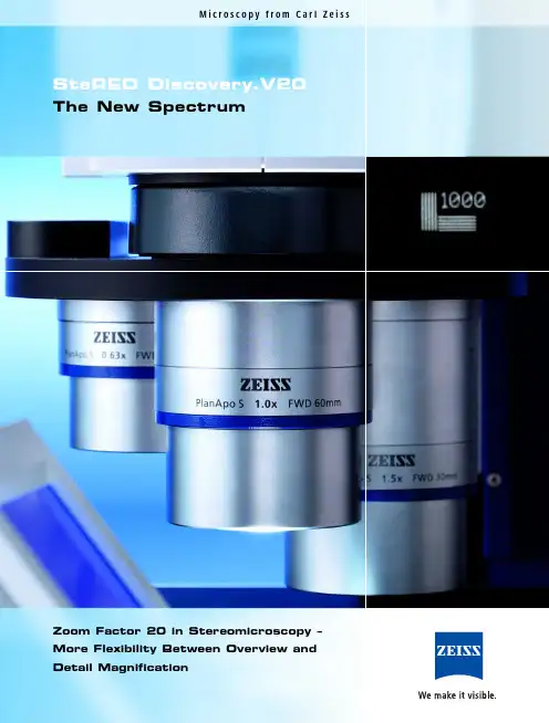
M i c r o s c o p y f r o m C a r l Z e i s sSteREO Discovery.V20The New SpectrumZoom Factor 20 in Stereomicroscopy –More Flexibility Between Overview andDetail MagnificationSFact Innovation:Never Before hasthe MagnificationSpectrum beenLarger.There’s a new performance standard in the demandingworld of stereomicroscopy: Zoom Factor 20. The factorfor the largest spectrum between overview and detailmagnification. The microscope: SteREO Discovery.V20.A Carl Zeiss design. And a research instrument withwhich the pioneer of the CMO principle (C ommon M ainO bjective) has once again broken new ground for thefuture of stereomicroscopy after the telescope principle.The development of the SteREO Discovery.V20 hasexceeded the limits of conventional modes of action.Founded on a new technological base and integratedinto the SteREO* generation series from Carl Zeiss,SteREO Discovery.V20 is highly impressive and boastsa superior performance profile. For maximum precisionand considerably more freedom in biology, medical andindustrial labs. The new features:• planapochromatic corrected microscope bodieswith a zoom range of 20:1• high end magnification of up to 345x(with eyepiece 10x)• maximum resolution of 1000LP/mm(with objective PlanApo S 2.3x)• excellent 3D-effect up to the highestmagnification• comfortable, securely reproducible operationaland control concept with SyCoP• seamless integration into the modular systemof the SteREO Discovery generation* SteREO – Stereomicroscopy Redefined in Ergonomics and Optics2teREO Discovery.V2034SyCoP 1.2.3.3.Extraordinarily large working area:the stand design with decentralized profile column S.The Performance Factor:Superiority Can be DocumentedAt the borders of technical possibilities details become a critical factor. Better optics is responsible for a visible improvement in image information. The easier operation concept delivers faster results. Fac-tors which Carl Zeiss places the utmost importance on in development and consistently optimizes until the peak of performance is reached. The results create new benchmarks. At any place where living objects or material samples are observed, manipulated or documented in detail, three-dimensionally and with high resolution or high contrast.1A in 3D: Spatial impressionWith SteREO Discovery.V20, higher magnifications can also be realized with smaller lenses thanks to the large zoom range of the microscope body. The smaller stereo angles associated improve the 3D impression of the microscopic image. The result:you remain more relaxed during observation and notice even the smallest details.Secure the highest magnifications: StabilityHigh image resolution and end magnification place new demands on the stability of the stand system of this stereomicroscope. All relevant components were designed and built according to the most modern methods. The stands feature a significantly higher rigidity and is considerably less susceptible to vibrations than previous systems. The motor focusing makes fine focusing in intervals of 350 nm in a range of 340 mm for loads of up to 17 kg possible.1.With SyCoP,even the most complex stereomicroscopic operation procedures can be handled comfortably.Without letting the sample out of your sight.With one hand,reliable and flawless.2.The new SteREO Discovery.V12 zoom body is parfocally adjusted.For pin sharp pictures in the complete magnification range.5Increase free space:Zoom factor 20:1The largest range from overview to detail –SteREO Discovery.V20 has brought a new zoom range into the research laboratory. And what’s more: even the basic configuration of this top ste-reomicroscope offers an end magnification of 150x.Equipped with the nosepiece S. cod as well asthe objectives PlanApo S 0.63x, PlanApo 1x and PlanApo S 2.3x, SteREO Discovery.V20 covers a magnification range from 4.7 to 345x. That is a factor of 73! With only one turn of the nosepiece.Decide economically:The SteREO Discovery upgrade conceptSteREO Discovery offers a wide spectrum of com-patible modules and accessory components. No matter what instrument type you choose, you have the freedom to upgrade your system according to your needs at any time. Up to the highest-capacity Imaging System that stereomicroscopy has to offer currently.Sophisticated and universally compatible:the wide accessory range fits for every instrument type of the SteREO Discovery generation.Intelligent operation: SyCoPSyCoP stands for S ystem C ontrol P anel and for a considerable gain in time, overview capability and flexibility in the operation of increasingly complex operation procedures. Designed especially for the demands of stereomicroscopy, the novel operation concept combines joystick, keys and touch screen in the handy design of a computer mouse. With SyCoP ,almost all important microscope functions can be controlled virtually location-independent. Fast, pre-cise and reproducible. Without removing your eye from the eyepiece ocular. Your attention stays on the object. In addition, SyCoP provides current data about the total magnification, object field, resolution and depth of focus of your microscope setting.SyCoP is an option for the future. New functions and further accessories are integrated through the open CAN-Bus concept.SteREO Discovery.The Technology Factor:Exceeding the LimitsAt the limits:The conventional technologyThe centerpiece of a CMO stereomicroscope is the pancrat (microscope or zoom body). During zoom-ing, lenses are moved and must be brought into a certain position in relation to other securely in-stalled lenses with extraordinary precision. Until now a mechanical curve – a simple metal piece milled with great care – determined largely parts the exactness of the traverse path of these lenses and thereby the overall microscope quality. The precision required for new stereomicroscope generations can no longer be fulfilled in this way. The solution:The new active principleOn the SteREO Discovery.V20, this mechanical curve has been replaced by a virtual one. The movable lenses are moved with a stepping motor and posi-tioned exactly with a processor. The microscopic images then stay considerably sharper. That has some definite advantages for your research applications:• 3D images can be viewed in the stereomi-croscope in a noticeably more relaxed way The partial images which are produced for our eyes are much sharper and better coordinated. The effort of the brain to create a 3D imageis less.• Sharper images producecontrast improvementsParticularly decisive when the stereomicroscopeis used in high and the highest magnifications. Microscopy pushed to the limits of useful magnification.• Higher magnifications with alarger zoom rangeUntil recently a zoom with a factor of 16 butnot higher was considered technically possible but this limit can now be exceeded considerably with this new technology. And it’s affordable.Carl Zeiss has created a new milestone in stereomi-croscopy with the SteREO Discovery.V20. Over 30 invention disclosures and patent applications are pro-viding that this technological advantage is preserved.Better 3D images, higher resolution, larger zoomranges – technologically, the conventional stereo-microscope has reached its limits. Each lens, eachmechanical detail exhibits tolerances – regardless ofthe precision of the production. The higher thedemands on resolution and magnification become,the less acceptable are these tolerances.Fast,flexible and effective: the final assembly of the stereomicroscopeSteREO Discovery in the clean rooms of Carl Zeiss Micro scopy GmbHin Jena.It is customized as “individual item chain production according tothe Wertstrom design criteria”.671.2.3.4.5.6.V20Zoom position of pancratD e f o c u s p o s i t i o n i n µmDepth of field curve, with in these parameters the picture is in focusTypical defocus curve of one channel with classical adjustment (pancrat with mechanical zoom curve)T ypical defocus curve of one channel of the SteREO Discovery.V20(pancrat with virtual electronically derived curve)1.Before the assembly begins each lens is exactly calibrated against a “0-lens type”.This lens value is digitally saved in a data pool – the basis on which computer-calculated combinations are established.By doing this,an optimally coordinated lens family is developed for every individual microscope2.Rotating reflexes of a lens.As soon as it is in the circle…3....a moveable micro clapper of the computer-controlled glue leveling machine autonomously undertakes the fine alignment.4.After being moved into place,the lenses are immediately fixed.Precision tools automatically lay high-precision,uninterrupted glue beads through a strong cannula 0.5 mm wide.5.Hardening of the glue beads under UV-radiation.6.In the pancrat adjusting device,the precise procedures of all moveable optical elements are programmed.To do this,around 7000 supporting points are analyzed via computer.In doing so,each stereomicroscope obtains its own correc-tion – its own individual zoom control curve.Illustration of the defocus curve of a classical mechanical pancrat in comparison to a motorized one (SteREO Discovery.V20).It is clear that the motorized pan-crat differs from the 0 line about half less than the mechanical one.That means:SteREO Discovery.V20 with motorized pancrat delivers twice as sharp images.8S1. 2. 4.3.1. Tubes Today ergonomics is a basic demand on microscopy.The user’s posture should remain relaxed even over long periods of work. An important factor for this are the observation tubes. The eyepiece sockets are swingable and adjustable in two levels. With the ergotube the angle of vision can be individually adjusted between 5 and 45 degrees.2. ObjectivesObjectives largely determine the image quality –and they are a relevant economic factor. The selec-tion of objectives for the SteREO Discovery.V20receives special attention for a reason. The spectrumranges from the cost-effective objectives of the Achromat series to the high-capacity Plan-Achromat objectives to the Plan-Apochromat series, which meets the highest requirements.3. StagesDesigned to move your objects gently and jolt-free during observation – a wide spectrum of different stages is available for the SteREO Discovery.V20.According to your needs, choose from sliding,rotating, mechanical or ball-and-socket stages.The motorized mechanical stage offers an addi-tional advantage in precision when adjusting and controlling objects: precisely accurate, fast and reproducible.4. FluorescencePentaFluar S is the retrofittable intermediate tube with a coaxial fluorescence mechanism which con-verts your SteREO Discovery.V20 into a high-capacity fluorescence system. The filter turret holds up to five filter modules and there are also many possibilities regarding illumination. Besides the well-established HBO lamps, X-Cite 120 with a liquid lightguide is recommended.The Flexibility Factor:the Upgrade Possiblities are EndlessThe modular construction of the SteREO Discovery.V20is typical for main lens stereomicroscopes. The multi-tude of accessory components which you can have installed on the high-capacity stereomicroscope in order to create an effective observation and docu-mentation system is correspondingly wide . The flexi-bility is unusual: completely integrated into the Carl Zeiss system world and equipped with intelligent interfaces, each component can be installed for every instrument type in the SteREO Discovery range.teREO Discovery.V205. 6.8.5. CamerasThe demands on the documentation of microscopic images in research are as different as the projects themselves. The spectrum of digital microscope cameras for the innovative high-performance system SteREO Discovery.V20 is correspondinglydiverse. Starting with digital consumer cameras through to professional cameras of the microscope camera family AxioCam, Carl Zeiss offers you a suit-able price and performance class for every demand.6. IlluminationThe quality of illumination essentially determines the quality of the results – in particular with stereo-microscopic contrasts. With an elaborate system of interfaces and adapters, SteREO Discovery.V20 can be equipped with modern fiber optic LED-compo-nents. Optimal for the illumination and contrasting of various objects.7. OperationCompletely motorized, SteREO Discovery.V20 offers reproducibility and considerable simplifications for your experiment procedures. In particular for con-trolling object details as well as for setting illumina-tion and contrasts. In addition, the innovative control system is now available. Designed to be user-friendly and securely operable. This is the foundation of thefact that the control of the current highest-capacity stereomicroscopy research device practically runsitself.8. Microscope SoftwareAxioVision is the superior software for microscopecontrol, image acquisition, image processing,image administration and archiving. With a univer-sal modular design and upgradeable according toyour needs from the basic version to the mostdemanding special configuration. The microscopesoftware from Carl Zeiss is completely integrated tothe current highest-capacity analysis platform andholds a top position worldwide on account of itssimple operation principle and its high productivity.9101.2.3.STrue to reality, completely shift-free 3D imaging of the researched specimen are the demands of contemporary stereomicroscopy. From a complete overview down to the smallest detail like organs,tissues and neurons. Now, Carl Zeiss raises the bar even higher.SteREO Discovery.V20 with its zoom of 20x doesn’t just deliver a large magnification range, it also shines with a brilliant image quality in the research of living specimen and other objects of Life Science. The con-sequent minimization of stray light of all tubes, the zoom body and the objectives as well as the indi-vidually tailored zoom curve, allow for a rich contrast in the images over the entire zoom range – from the overview up to the highest magnification. The large base and the grand front lens add to the unique 3D effect.Therefore the SteREO Discovery.V20 ensures an image quality that you can count on in research facilities and laboratories of biology and medicine. It is also ideal to observe and research model organ-isms of developmental biology.Practical Life Sciences:High Magnifications,Excellent Depth Perception1.Mouse embryo,stained,transmitted-light brightfield,objective PlanApo S 0.63x,magnification 4.7x*2.Mouse embryo,stained,transmitted-light brightfield,objective PlanApo S 0.63x,magnification 94x*3.Diatome,transmitted-light darkfield,objective PlanApo S 2.3x,magnification 345x*111.2.teREO Discovery.V20The demands for high-end stereomicroscopes rise with the perception of smaller details. On one hand there is the need for fast orientation of where you are in the specimen and that requires a large overview. On the other hand there is the need for observing and documenting the smallest detail in a rapid switch from the overview image – ideally without re-focussing.The SteREO Discovery.V20 with a zoom of 20x gives you a vast advantage in your lab. It is the only stereomicroscope that allows a fast switch from overview to detail image. Here the motorised zoom delivers a precise and free to choose zoom position.And it only differs less than 1%. That means a reproducibility of more than 99%! This is the pre-cision of an ideal research instrument with its always correctly scaled images to measure and document tasks in micromechanics and quality control. And it is a safe investment into a new dimension of achievement.Practical Materials:99% Reproducibility,100% Investment Security1.Semiconductor,reflected-light darkfield,objective PlanApo S 1x,magnification 7.5x*2.Semiconductor,reflected-light darkfield,objective PlanApo S 1x,magnification 150x*3.Semiconductor,reflected-light darkfield,objective PlanApo S 1x,magnification 20x*,Extended Depth of Focus* Total magnification through eyepiecesObjectivesEyepiecesSteREO Discovery.V20:The Technical DataCarl Zeiss Micro scopy GmbH 07745 Jena, Germany ********************www.zeiss.de/stereo-discoveryP r i n t e d o n e n v i r o n m e n t a l l y -f r i e n d l y p a p e r , b l e a c h e d w i t h o u t t h e u s e o f c h l o r i n e .S u b j e c t t o c h a n g e .46-0128 e 05.2007。

芯片检测光谱
芯片检测中的光谱技术是一种利用物质对特定波长光的吸收、发射或反射特性来进行材料分析的方法。
光谱检测技术在半导体制造和芯片质量控制中扮演着重要角色,它可以提供关于材料成分、纯度、晶体结构以及表面缺陷等的信息。
以下是几种常用的光谱检测技术及其在芯片检测中的应用:
1. 傅里叶变换红外光谱(FTIR): FTIR可以用来检测芯片材料中的化学成分和分子结构。
通过分析材料对红外光的吸收光谱,可以识别出特定的官能团,进而判断出材料的类型和纯度。
2. 拉曼光谱: 拉曼光谱是一种无损检测技术,它通过分析光子散射后的能量变化来获得材料的分子振动信息。
拉曼光谱能够提供晶体结构、应力状态、掺杂水平等的信息,对于半导体材料的质量控制非常有用。
3. 紫外-可见光谱(UV-Vis): UV-Vis光谱可以用来评估芯片表面的光吸收特性,从而检测污染物、氧化层厚度以及其他表面特性。
4. 荧光光谱: 荧光光谱技术可以用来检测芯片上的某些元素或化合物,因为它们在受到激发后会发出特定波长的光。
这种技术对于检测芯片上的微量杂质特别有效。
5. 近红外光谱(NIR): NIR光谱检测通常用于测量材料的浓度和组成,因为许多化学物质在近红外区域有特定的吸收峰。
光谱检测技术的关键优势在于其非接触性和高灵敏度,这使得它们能够在不损坏芯片的情况下提供精确的材料属性信息。
此外,光谱技术通常能够快速进行,适合在线监测和实时质量控制。
随着光谱仪器的不断进步,它们在芯片制造和检测中的应用也在不断扩大,有助于提高芯片的生产效率和产品质量。
电感耦合等离子体发射光谱法的英文简称全文共3篇示例,供读者参考篇1Inductively Coupled Plasma Optical Emission Spectroscopy (ICP-OES) is a powerful analytical technique used in many scientific fields. This technique utilizes the high temperature of a plasma to atomize and excite samples for elemental analysis. ICP-OES provides high sensitivity, accuracy, and precision, making it a popular choice for analyzing trace elements in various sample types.The process of ICP-OES involves generating a plasma by applying a high-frequency radio frequency (RF) current to a flowing gas, typically argon. The intense heat of the plasma vaporizes the sample and excites the atoms to emit characteristic light at specific wavelengths. This emitted light is then dispersed by a spectrometer and detected by a charged-coupled device (CCD) detector. The intensity of the emitted light is proportional to the concentration of the element in the sample, allowing for quantitative analysis.ICP-OES is widely used in environmental monitoring, pharmaceutical analysis, forensic science, and materials science, among other areas. It can detect a wide range of elements, from alkali metals to rare earth elements, with detection limits as low as parts per billion. Additionally, ICP-OES can analyze multiple elements simultaneously, making it a fast and efficient tool for elemental analysis.Overall, ICP-OES is a versatile and reliable technique for elemental analysis, providing accurate and precise results for a wide range of sample types. Its high sensitivity and ability to analyze multiple elements simultaneously make it an essential tool in many research and industrial laboratories.篇2Title: ICP-OES: The Technique Behind Inductively Coupled Plasma Optical Emission SpectroscopyIntroductionInductively Coupled Plasma Optical Emission Spectroscopy, commonly abbreviated as ICP-OES, is a powerful analytical technique used for the quantitative analysis of elements present in a sample. This technique utilizes the principles of inductively coupled plasma (ICP) and optical emission spectroscopy (OES) toprovide accurate and precise measurements of the elemental composition of a sample. In this article, we will explore the fundamentals of ICP-OES and its applications in various fields.Principles of ICP-OESICP-OES operates by generating a high-temperature plasma consisting of ionized gas atoms by introducing a sample into an argon gas stream. The plasma is sustained by an induction coil, which induces an electric current that generates heat, forming a high-energy environment capable of atomizing and ionizing the sample components. As the atoms and ions return to their ground state, they emit light at characteristic wavelengths, which can be measured by a spectrometer to identify and quantify the elements present in the sample.Advantages of ICP-OESICP-OES offers several advantages over other analytical techniques, making it a preferred choice for elemental analysis in various industries. Some of the key advantages of ICP-OES include:- High sensitivity and detection limits: ICP-OES can detect elements at trace levels, making it suitable for a wide range ofapplications, including environmental monitoring and pharmaceutical analysis.- Multi-element analysis: ICP-OES is capable of analyzing multiple elements simultaneously, providing comprehensive information on the elemental composition of a sample.- Wide dynamic range: ICP-OES can analyze elements across a wide concentration range, from parts-per-billion to percent levels, making it suitable for diverse sample types.- Speed and efficiency: ICP-OES offers rapid analysis times, allowing for high sample throughput and increased productivity.- Minimal sample preparation: ICP-OES requires minimal sample preparation, saving time and reducing the risk of sample contamination.Applications of ICP-OESICP-OES is widely used in various industries and research fields for elemental analysis due to its versatility and accuracy. Some common applications of ICP-OES include:- Environmental analysis: ICP-OES is used for the analysis of trace elements in soil, water, and air samples to assess environmental contamination levels.- Geological analysis: ICP-OES is employed in the analysis of rocks, minerals, and ores to determine their elemental composition and identify valuable mineral deposits.- Pharmaceutical analysis: ICP-OES is used in the pharmaceutical industry for the analysis of drug formulations, determining the elemental impurities present in pharmaceutical products.- Food and beverage analysis: ICP-OES is utilized for the analysis of food and beverage products to ensure compliance with regulatory standards and assess product safety.ConclusionICP-OES is a versatile and reliable technique for elemental analysis, offering high sensitivity, multi-element capabilities, and rapid analysis times. With its wide range of applications in various fields, ICP-OES has become an essential tool for researchers, analysts, and industry professionals seeking accurate and precise elemental analysis. As technology continues to advance, ICP-OES is expected to play a key role in shaping the future of analytical chemistry and elemental analysis.篇3Inductively Coupled Plasma Emission Spectroscopy (ICP-ES) is a powerful analytical technique widely used in various fields including environmental monitoring, pharmaceutical analysis, and material science. This technique is based on the inductively coupled plasma (ICP) as the excitation source and the emission spectroscopy for detecting and quantifying elements present in a sample.ICP-ES offers several advantages over other analytical methods. Firstly, it provides a high sensitivity, allowing for the detection of trace elements at parts per billion or even parts per trillion levels. This makes ICP-ES ideal for analyzing samples with low concentrations of elements of interest. Secondly, ICP-ES has a wide dynamic range, enabling the simultaneous analysis of multiple elements present in a sample. This feature is particularly useful when analyzing complex samples containing a diverse range of elements. Additionally, ICP-ES offers excellent precision and accuracy, making it a reliable technique for quantitative analysis.The principle of ICP-ES involves the generation of ahigh-temperature plasma by inducing an electric current in a gas (typically argon) using a radiofrequency source. The plasma reaches temperatures of up to 10,000 Kelvin, causing the sampleto be atomized and ionized. As a result, the atoms and ions emit characteristic radiation when transitioning from excited states to ground states. The emitted radiation is then dispersed and detected by a spectrometer, allowing for the identification and quantification of elements based on their unique emission spectra.The use of inductively coupled plasma as the excitation source offers several advantages over other excitation sources, such as flame atomic absorption spectroscopy and graphite furnace atomic absorption spectroscopy. Firstly, the high temperature of the plasma ensures complete atomization and ionization of the sample, leading to higher sensitivity and lower detection limits. Secondly, the plasma provides a stable and robust excitation source, resulting in reliable and reproducible analytical results. Additionally, the high energy density of the plasma allows for the analysis of refractory elements that are difficult to atomize using other excitation sources.ICP-ES is a versatile technique that can be used for the analysis of a wide range of samples, including liquids, solids, and gases. It is commonly used for the analysis of environmental samples, such as water, soil, and air, to monitor the levels of toxic elements and pollutants. In the pharmaceutical industry, ICP-ESis used for the analysis of drug formulations to ensure compliance with regulatory standards. In material science, ICP-ES is employed for the analysis of metals, alloys, and ceramics to determine their elemental composition and purity.In conclusion, Inductively Coupled Plasma Emission Spectroscopy (ICP-ES) is a powerful analytical technique that offers high sensitivity, wide dynamic range, and excellent precision for the analysis of trace elements in various samples. Its use of inductively coupled plasma as the excitation source provides several advantages over other excitation sources, making it a popular choice in analytical laboratories worldwide. With its versatility and reliability, ICP-ES is a valuable tool for research, quality control, and environmental monitoring applications.。
山东化工SHANDONGCHEMICALINDUSTRY-158-2020年第49卷核磁共振波谱课程教学探索李晓虹(苏州大学材料与化学化工学部,江苏苏州215123)摘要:核磁共振波谱作为鉴定化合物结构、组分含量、动力学参数等信息的重要手段,在化学、医药、材料等领域科研生产中起着关键作用。
其课程教学长期以来受到理论内容难、仪器开放难等因素困扰’结合苏州大学核磁共振波谱课程的双语教学实践提出了相应的对策与改进举措,探讨通过更新改进教学方法和内容,突破传统教学模式,使学生从理论联系实践,从“会用”到“用好”核磁技术’关键词:核磁共振波谱;远程虚拟终端%网络课堂中图分类号:G642O文献标识码:B文章编号:1008-021X(2020)23-0158-02Exploration of Teaching in Nuclear Magnetic Resonance Spectroscopy CourseLi Xiaohong(Colleae of Chemist—,Chemicai Enginee/ng and Materials Science of Soochow University,Suzhou215123,China) Abstract:Nuclear magnetic resonance spectroscopy(NMR),as an Onportant method of studying compound structures, component contents and kinetic parameters,plays a key rolo in the fields of chemist—,pharmaceutical indust—and materials science.For a long time,its course teaching has been troubled by the dOficulta of theo—tical content and the lack of instmmentai peacicce.Based on ihebcocnguaoieachcngpeacicceooNMR couesecn Soochow Unceeesciy,ihcspapeedcscu s eshow iobeeak iheough iheieadciconaoieachcngmodebycmpeoecngiheieachcngmeihodsand conienis,soihaisiudeniscan combcneiheoeywcih peacicceand makegood useooNMRiechnooogy.Key wordt:NMR%VNC%online coa s es核磁共振波谱作为鉴定化合物结构的重要手段,对样品无损,分辨率高,较灵敏,可获得准确的定性定量信息。
钱玉,刘帅,金龙,等. 基于表面增强拉曼光谱的巴旦木氧化程度快速检测[J]. 食品工业科技,2023,44(24):286−293. doi:10.13386/j.issn1002-0306.2023020173QIAN Yu, LIU Shuai, JIN Long, et al. Rapid Detection of the Oxidation of Almonds Based on Surface Enhanced Raman Spectroscopy[J]. Science and Technology of Food Industry, 2023, 44(24): 286−293. (in Chinese with English abstract). doi:10.13386/j.issn1002-0306.2023020173· 分析检测 ·基于表面增强拉曼光谱的巴旦木氧化程度快速检测钱 玉1,刘 帅1,金 龙2,孙 美2,颜 玲1,刘长虹1,董保磊1, *,郑 磊1,*(1.合肥工业大学食品与生物工程学院,安徽合肥 230031;2.洽洽食品股份有限公司,安徽合肥 230031)摘 要:氧化程度对巴旦木营养和品质具有重要的影响,本研究的目的是建立一种灵敏、可靠的巴旦木氧化程度快速检测方法。
本研究首先通过表面配体交换的转相策略实现了水溶液中分散的金纳米粒子(AuNPs )快速、简便地向非极性的甲苯溶液中的转相。
UV-Vis 和透射电镜等表征结果表明转相后的AuNPs 的纳米形貌未发生明显的变化,可成功作为表面增强拉曼光谱(SERS )基底用于巴旦木油脂氧化程度的检测。
结果表明,巴旦木油脂位于1655 cm −1处的顺式双键的特征拉曼信号在氧化过程中逐渐减弱;选择酯键的1747 cm −1作为参比信号,其特征峰的相对强度I 1655/I 1747值与巴旦木的加速氧化时间呈良好线性关系(R 2=0.98),SERS 光谱结果结合主成分分析法可以用于实际巴旦木样品氧化程度的快速判定和分类。
近红外光谱法英文Near-Infrared SpectroscopyNear-infrared spectroscopy (NIRS) is a powerful analytical technique that has gained widespread recognition in various scientific and industrial fields. This non-invasive method utilizes the near-infrared region of the electromagnetic spectrum, typically ranging from 700 to 2500 nanometers (nm), to obtain valuable information about the chemical and physical properties of materials. The versatility of NIRS has led to its application in a diverse array of industries, including agriculture, pharmaceuticals, food processing, and environmental monitoring.One of the primary advantages of NIRS is its ability to provide rapid and accurate analysis without the need for extensive sample preparation. Unlike traditional analytical methods, which often require complex sample extraction and processing, NIRS can analyze samples in their natural state, allowing for real-time monitoring and decision-making. This efficiency and non-destructive nature make NIRS an attractive choice for applications where speed and preservation of sample integrity are crucial.In the field of agriculture, NIRS has become an invaluable tool for the assessment of crop quality and the optimization of farming practices. By analyzing the near-infrared spectra of plant materials, researchers can determine the content of various nutrients, such as protein, carbohydrates, and moisture, as well as the presence of contaminants or adulterants. This information can be used to guide precision farming techniques, optimize fertilizer application, and ensure the quality and safety of agricultural products.The pharmaceutical industry has also embraced the use of NIRS for a wide range of applications. In drug development, NIRS can be used to monitor the manufacturing process, ensuring the consistent quality and purity of active pharmaceutical ingredients (APIs) and finished products. Additionally, NIRS can be employed in the analysis of tablet coatings, the detection of counterfeit drugs, and the evaluation of drug stability during storage.The food processing industry has been another significant beneficiary of NIRS technology. By analyzing the near-infrared spectra of food samples, manufacturers can assess parameters such as fat, protein, and moisture content, as well as the presence of adulterants or contaminants. This information is crucial for ensuring product quality, optimizing production processes, and meeting regulatory standards. NIRS has been particularly useful in the analysis of dairy products, grains, and meat, where rapid and non-destructive testing is highly desirable.In the field of environmental monitoring, NIRS has found applications in the analysis of soil and water samples. By examining the near-infrared spectra of these materials, researchers can obtain information about the presence and concentration of various organic and inorganic compounds, including pollutants, nutrients, and heavy metals. This knowledge can be used to inform decision-making in areas such as soil management, water treatment, and environmental remediation.The success of NIRS in these diverse applications can be attributed to several key factors. Firstly, the near-infrared region of the electromagnetic spectrum is sensitive to a wide range of molecular vibrations, allowing for the detection and quantification of a variety of chemical compounds. Additionally, the ability of NIRS to analyze samples non-destructively and with minimal sample preparation has made it an attractive choice for in-situ and real-time monitoring applications.Furthermore, the development of advanced data analysis techniques, such as multivariate analysis and chemometrics, has significantly enhanced the capabilities of NIRS. These methods enable the extraction of meaningful information from the complex near-infrared spectra, allowing for the accurate prediction of sample propertiesand the identification of subtle chemical and physical changes.As technology continues to evolve, the future of NIRS looks increasingly promising. Advancements in sensor design, data processing algorithms, and portable instrumentation are expected to expand the reach of this analytical technique, making it more accessible and applicable across a wider range of industries and research fields.In conclusion, near-infrared spectroscopy is a versatile and powerful analytical tool that has transformed the way we approach various scientific and industrial challenges. Its ability to provide rapid, non-invasive, and accurate analysis has made it an indispensable technology in fields ranging from agriculture and pharmaceuticals to food processing and environmental monitoring. As the field of NIRS continues to evolve, it is poised to play an increasingly crucial role in driving innovation and advancing our understanding of the world around us.。
微区光谱英文Micro-area spectroscopy is an important analytical technique that allows researchers to obtain detailed information about the chemical and physical properties of small areas or points on the surface of a material. This technique involves the use of a focused beam of light or charged particles to excite the sample and then detect the emitted or scattered radiation. This document will provide an overview of micro-area spectroscopy, including its applications, advantages, and limitations.Applications of Micro-Area SpectroscopyMicro-area spectroscopy has a wide range of applications in various fields such as materials science, biology, and environmental science. Some of the most common applications of micro-area spectroscopy are as follows:1. Imaging of Microstructures: Micro-area spectroscopy can reveal the chemical composition and morphology of various microstructures on a material surface. For example, it can be used to map the distribution of specific elements orcompounds in a sample or to image the topography ofa surface with high spatial resolution.2. Characterization of Thin Films: Micro-area spectroscopy can be used to characterize the thickness, composition, and optical properties of thin films. This is useful in the development of new materials and in quality control of finished products.3. Analysis of Biological Samples: Micro-area spectroscopy can provide information about the chemical composition and structure of biological samples, such as tissues, cells, and biomolecules. This information can be used to study disease processes, drug interactions, and cellular activities.4. Environmental Analysis: Micro-area spectroscopy can be used to identify and quantify the presence of pollutants in soil, water, and air. This information can help in the monitoring and remediation of contaminated environments.Advantages of Micro-Area SpectroscopyThere are several advantages of micro-area spectroscopy that make it a popular technique for many researchers:1. Increased Sensitivity: Micro-area spectroscopy can detect very small amounts of materials or compounds, which is important for the analysis of small or trace samples.2. High Spatial Resolution: Micro-area spectroscopy can obtain detailed information about the chemical and physical properties of small areas or points on a sample surface.3. Non-Destructive Analysis: Micro-area spectroscopy is a non-destructive technique that can be used to analyze samples without altering or damaging them.4. Versatility: Micro-area spectroscopy can be used with a wide range of samples, including opaque, transparent, or reflective materials.Limitations of Micro-Area SpectroscopyMicro-area spectroscopy also has some limitations that should be considered when using this technique for analysis:1. Time-Consuming: Obtaining high-quality spectra using micro-area spectroscopy can be time-consuming, especially for samples that require long acquisition times.2. Sample Preparation: Samples must becarefully prepared to ensure that they are suitable for micro-area spectroscopy. This can be a complex and time-consuming process.3. Cost: Micro-area spectroscopy requires expensive equipment and specialized expertise, which can make it cost-prohibitive for some laboratories.ConclusionIn conclusion, micro-area spectroscopy is a valuable technique for the analysis of small samples in a wide range of applications. Its high sensitivity and spatial resolution make it an important tool for researchers working in materials science, biology, and environmental science. However, the limitations of micro-area spectroscopy should be carefully considered before it is usedfor analysis.。
NASA's Alice ultraviolet (UV) spectrograph aboard the European Space Agency's Rosetta comet orbiter has delivered its first scientific discoveries. Rosetta, in orbit around comet 67P/Churyumov-Gerasimenko, is the first spacecraft to study a cometup close. As Alice began mapping the comet's surface last month, it made the first far ultraviolet spectra1 of a cometary surface. From these data, the Alice team discovered that the comet is unusually dark at ultraviolet wavelengths2 and that the comet's surface -- so far -- shows no large water-ice patches. Alice is also already detecting both hydrogen and oxygen in the comet's coma3, or atmosphere."We're a bit surprised at both just how very unreflective the comet's surface is, and what little evidence of exposed water-ice it shows," says Dr. Alan Stern, Alice principal investigator4 and an associate vice5 president of the Southwest Research Institute (SwRI) Space Science and Engineering Division.Developed by SwRI, Alice is probing the origin, composition and workings of the comet, gaining sensitive, high-resolution compositional insights that cannot be obtained by either ground-based or Earth-orbital observations. The ultraviolet wavelengths Alice observes contain unique information about the composition of the comet's atmosphere and the properties of its surface."As the mission progresses, we will continue to search for surface ice patches and ultraviolet color and composition variations across the surface of the comet," says Dr. Lori Feaga, Alice co-investigator at the University of Maryland.Alice is one of three instruments funded by NASA flying aboard Rosetta. Alice has more than 1,000 times the data-gathering capability6 of instruments flown a generation ago, yet it weighs less than 4 kilograms and draws just 4 watts7 of power.A sister Alice instrument was developed by SwRI and was launched aboard the New Horizons spacecraft to Pluto8 in January 2006 to study that distant world's atmosphere. It will reach Pluto in July 2015. SwRI also built and operates Rosetta's Ion and Electron Spectrograph (IES), another instrument with miniaturized electronic systems. With a mass of 1.04 kilograms, IES achieves sensitivity comparable to instruments weighing five times more.To reach its comet target, the Rosetta spacecraft executed four gravity assists (three from Earth, one from Mars) and a nearly three-year period of deep space hibernation9, waking up in January 2014 in time to prepare for its rendezvous10 with Churyumov-Gerasimenko. Rosetta also carries a lander, Philae, that will drop to the comet's surface in November 2014, attempting the first-ever direct observations of a comet surface.Rosetta is an ESA mission with contributions from its member states and NASA. Rosetta's Philae lander is provided by a consortium led by DLR, MPS, CNES and ASI.Airbus Defense11 and Space built the Rosetta spacecraft. NASA's Jet Propulsion Laboratory (JPL) manages the U.S. contribution of the Rosetta mission for NASA's Science Mission Directorate in Washington, under a contract with the CaliforniaInstitute of Technology (Caltech). JPL also built the Microwave Instrument for the Rosetta Orbiter and hosts its principal investigator, Dr. Samuel Gulkis. SwRI (San Antonio and Boulder12, Colo.) developed the Rosetta orbiter's Ion and Electron Sensor13 and Alice instrument and hosts their principal investigators14, Dr. James Burch (IES) and Dr. Alan Stern (Alice).词汇表:1 spectran.光谱参考例句:The infra-red spectra of quinones present a number of interesting features. 醌类的红外光谱具有一些有趣的性质。
冷原子光谱法英语Okay, here's a piece of writing on cold atom spectroscopy in an informal, conversational, and varied English style:Hey, you know what's fascinating? Cold atom spectroscopy! It's this crazy technique where you chill atoms down to near absolute zero and study their light emissions. It's like you're looking at the universe in a whole new way.Just imagine, you've got these tiny particles, frozen in place almost, and they're still putting out this beautiful light. It's kind of like looking at a fireworks display in a snow globe. The colors and patterns are incredible.The thing about cold atoms is that they're so slow-moving, it's easier to measure their properties. You can get really precise data on things like energy levels andtransitions. It's like having a super-high-resolution microscope for the quantum world.So, why do we bother with all this? Well, it turns out that cold atom spectroscopy has tons of applications. From building better sensors to understanding the fundamental laws of nature, it's a powerful tool. It's like having a key that unlocks secrets of the universe.And the coolest part? It's just so darn cool! I mean, chilling atoms to near absolute zero? That's crazy science fiction stuff, right?。
专利名称:Spectroscopic microscopy with image-drivenanalysis发明人:Federico Izzia,Kathleen J.Schulting,Alexander Grenov申请号:US11846499申请日:20070828公开号:US07496220B2公开日:20090224专利内容由知识产权出版社提供专利附图:摘要:In a spectroscopic microscope, a video image of a specimen is analyzed to identify regions having different appearances, and thus presumptively differentproperties. The sizes and locations of the identified regions are then used to position the specimen to align each region with an aperture, and to set the aperture to a size appropriate for collecting a spectrum from the region in question. The spectra can then be analyzed to identify the substances present within each region of the specimen. Information on the identified substances can then be presented to the user along with the image of the specimen.申请人:Federico Izzia,Kathleen J. Schulting,Alexander Grenov地址:Middleton WI US,Oregon WI US,Madison WI US国籍:US,US,US代理人:Charles B. Katz,Michael C. Staggs更多信息请下载全文后查看。
Spectromicroscopy of interfacial interactions betweenthin Ni ®lms and a Au±Si surfaceL.Gregoratti a ,A.Barinov b ,L.Casalis a ,M.Kiskinova a,*aSincrotrone Trieste,Area Science Park Basovizza,34012Trieste,Italy bKurchatov Institute,Kurchatov sq.1,123182Moscow,RussiaReceived 29May 2000;accepted 24August 2000AbstractThe interaction between a (p 3Âp3)R308-Au/Si surface with thin ( 2ML)Ni ®lms with different dimensions deposited at 300K is studied by means of a scanning photoelectron spectromicroscopy.The interfacial processes occurring at different Ni coverages and annealing temperatures are probed with lateral resolution of 0.12m m.The results have revealed that Ni displaces Au from the Si surface and the presence of Au lowers the formation temperature of the Ni disilicide phase.It has been found that the behavior of the Ni/Au±Si interfaces at high temperatures,in particular the changes in the morphology of the interface and the formation of well de®ned Ni x (Au 1Àx )Si 2islands,is strongly in¯uenced by the dimensions of the Ni patches.The observed evolution of the Ni/Au±Si interfaces is interpreted considering the high solubility of Ni in Si,the miscibility between Ni and Au and the Au±Si alloying at elevated temperatures.#2001Elsevier Science B.V .All rights reserved.Keywords:Silicide interfaces;Surface structures;Spectromicroscopy;Surface reactions;Si;Ni;Au1.IntroductionTransition metal silicides have widely been used in microelectronic technology as Schottky barriers,con-tacts,interconnects,etc.In very large scale integration circuits the diminishing widths of the metal silicide lines can lead to undesirable increase of the resistance.The resistance of Ni silicides turned out to be the least affected by the shrinking dimensions of the line widths which makes them attractive for potential applications [1].Among the various nickel silicide phases the NiSi 2is the most desired but its nucleation-controlled for-mation requires a high temperature.However,asshown recently,even after high temperature annealing NiSi still coexists with NiSi 2[2,3].Recent structural and Rutherford backscattering spectroscopy studies reported that the presence of Au could modify the solid state reaction between Ni and Si lowering the nucleation temperature of NiSi 2[4,5].Earlier studies of formation of other transition metal silicides in thepresence of an Au interlayer of thickness >100AÊrelated the effect of Au to enhanced Si out-diffusion [6].However,the mechanism of the Au `promotion'effect is not clear.It is well known that depending on the temperature a Au±Si alloy or ordered 2D phases mixed with agglomerated Au islands can dominate the Au/Si interface [7,8].In the case of bimetallic sys-tems,the evolution of the interface becomes more complicated because it depends on many factors,such as metal±Si af®nity,miscibility of the metals withtheApplied Surface Science 171(2001)265±274*Corresponding author.Tel.: 39-040-375-85-49;fax: 39-040-375-85-65.E-mail address :kiskinova@elettra.trieste.it (M.Kiskinova).0169-4332/01/$±see front matter #2001Elsevier Science B.V .All rights reserved.PII:S 0169-4332(00)00763-7formed phases,etc.[9].Very important also is the surface morphology of thin NiSi 2®lms grown in the presence of Au and the lateral distribution of Au during temperature treatments of the bimetallic interface.The aim of the present study is to get insight of the interactions between very thin Ni ®lms and Si in the presence of Au,using a method that combines surface chemical sensitivity with high spatial resolution.Par-ticular attention is paid to the changes in the chemical state and lateral distribution of Au and Ni.Here,we started with a very well de®ned two-dimensional (2D)(p 3Âp 3)R308-Au ordered phase on Si(111)and studied the interfacial reactions with Ni ®lms,depos-ited through masks of different dimensions.The con-centration pro®le across the edge of such `con®ned'Ni ®lms allows investigation of the effect of the ®lm thickness under the same experimental conditions and of the mass transport of material during temperature treatments.Essential for better understanding the behavior of this bimetallic interface is the possibility of reference measurements of the Ni-free H Au/Si region outside the Ni patch.It should also be noted that the morphology of the `con®ned'®lms is similar to interconnect and contacts in the electronic devices,where mass transport phenomena and edge effects may become important.Here,we will show that the processes dominating the thermal evolution of the Ni/Au±Si interface depend on the actual size of the Ni patches with respect to the substrate dimensions.This con®rms that the con®ned metal ®lms can behave differently from discontinuous ®lms,an important issue in characterization of metal/semiconductor interfaces at length scales of interconnects and contacts.2.ExperimentalAll experiments were carried out with the scanning photoelectron microscope (SPEM)at the ELETTRA synchrotron light source,which uses zone plate optics for producing a microprobe with sub-micrometer dimensions [10].Here we used a zone plate,which provided spatial resolution of 0.12m m.The micro-scope works in an imaging mode with the electron analyzer collecting photoelectrons with a speci®c kinetic energy,while scanning the sample with respectto the beam,or in a micro-spectroscopy mode,mea-suring photoelectron (PE)spectra from a feature selected from the image.The measurement station is equipped with facilities for specimen preparation,metal evaporation and sample characterization by low energy electron diffraction (LEED)and Auger elec-tron spectroscopy (AES).The temperature of the samples is controlled using an optical pyrometer with calibrated emissivity factors.The initial Au/Si interface was prepared by anneal-ing of about 1ML Au ®lm deposited on a (7Â7)-Si(111)(n-doped,0.17O cm)sample with dimen-sions 3mm Â7mm.1ML equals the (1Â1)-Si(111)surface atomic density,7X 8Â1014atoms/cm 2.The LEED,AES and PE spectra indicated that the formed (p 3Âp 3)R308structure contains 0X 9Æ0X 1ML Au and consists of uniform H 3-domains and domain walls [11,12].1and 2ML of Ni were post-deposited through masks with dimension 1X 4mm Â7mm and 0X 6mm Â0X 1mm.In the former case,bout 43%of the H 3-Au/Si surface was covered by Ni,whereas the smaller Ni patch covered only 0.3%of the sample.During all evaporation and annealing procedures the pressure was kept in the low 10À10mbar range.All measurements were per-formed with a photon energy of 493eV and energy resolution of 0.3eV.For the used photon energy the effective escape depths of Si 2p,Au 4f and Ni 3p photoelectrons for our geometry are estimated to bearound 4AÊ.The Fermi energy position is determined from the valence band spectrum of the fresh Ni ®lm deposited on the Ta clips.Doniach±Sunjic functions convoluted with gaussians,which simulated the over-all energy spread,are used for ®tting the PE spectra [13].3.ResultsThe Ni 3p and Au 4f maps and the valence band (VB),Si 2p,Au 4f and Ni 3p spectra,measured inside,outside and across the edge of the Ni ®lms,are used as ®ngerprints for the evolution of the interface as a function of the Ni coverage and temperature.Fig.1(a)shows a typical Ni 3p map,where the part covered with Ni appears bright.In the present study,the concentration pro®le measured across the edge of the Ni ®lm varies between 0and 2ML and extends266L.Gregoratti et al./Applied Surface Science 171(2001)265±274over about 15m m,as can be seen in Fig.1.This allows precise monitoring of the Ni coverage effects.As will be demonstrated in the next sections the lateral changes in the contrast of the Ni 3p and Au 4f maps inside the patch re¯ect the temperature-induced changes in the morphology of the interface.3.1.Coadsorption of Ni at 300KFigs.1(b)and 2show selected VB,Ni 3p,Au 4f and Si 2p spectra taken on the Ni-free H 3-Au/Si area and across the edge of a 2ML Ni ®lm deposited at 300K.The VB spectrum of the H 3-Au/Si surface is domi-nated by the Au 5d states,forming the doublet below À4eV in Fig.1(b).The intensity close to the Fermi level increases with increasing Ni coverage with a maximum moving from approx.À2.0to À1.6eV.TheNi 3p panel in Fig.1(b)shows that the Ni 3p intensity gradually increases with Ni coverage and shifts by 0.6eV in the coverage range 0.8±2.0ML,reaching the energy position of the metallic Ni state (À67.0eV).The Au 4f spectra of the H 3-Au/Si phase,shown in Fig.2,are shifted by À1.3eV with respect to the metallic Au energy position and require a larger gaussian width,most likely due to the different bond-ing con®gurations within the domains and the domain walls.The Si 2p spectra of the H 3-Au/Si phase are ®tted using a bulk component,B-Si,and two `reacted'Si components,R1and R2.The B-Si 2p 3/2energy,À99.4eV ,coincides with the B-Si position of the pinned (7Â7)-Si(111)surface,indicating negligible band-bending effects.The R1-Si and R2-Si compo-nents are chemically shifted by À0.3and À0.5eV with respect to the B-Si component.They are assigned to the Si atoms bonded to Au within the H 3-domains (R1)and at the Au-rich domain walls (R2).The R1/R2ratio depends on the density of the domain walls,which can cover up to 34%of the surface [14].The Si 2p and Au 4f spectra undergo intensity and lineshape changes with increasing Ni coverage,which are man-ifested by the spectra in Fig.2and the plots in Fig.3(a).The Au 4f spectra become broader in the Ni coverage range 0.9±1.5ML and sharpen with further increase of the Ni coverage.Close to 2ML the Au 4f lineshape and energy position (À84.1eV)become practically identical with that of the metallic Au state.The metallic Au component appears and grows at the expense of the `reacted'Au component at Ni coverage !0.8ML,which causes the broadening of the Au 4f peaks up to 1.5ML.The change of the Au chemical state at high Ni coverages also leads to a shift of the Au 5d states (Fig.1)by 0.3eV .In the Ni coverage range 0±1.0ML,the intensity of the Si 2p signal decreases due to attenuation of the B-Si and R1components.The ®ts of the Si 2p spectra in Fig.2reveal that 0.25ML a new R3-Si component grows rapidly.From here on we will use the assignment R3-Si for the Si atoms with a mixed Ni Au coordination.It should be noted that the binding energy of the growing R3-Si at 300K is almost identical to the R2component assigned to the Si coordinated with Au at the domain walls of the H 3-Au structure,where the local Au coverage is far beyond 1ML.This assigns the R2-Si and R3-Si as components corresponding to Si atoms in a metal-rich environment.As can be seen in Fig.3(a)theR3-SiFig.1.(a)Ni 3p 64Â8m m 2map centered close to the edge of a 2ML Ni patch at 300K and the corresponding Ni concentration pro®le across the edge;(b)valence band (left)and Ni 3p (right)spectra as a function of the local Ni coverage.The spectra are taken in spots across the patch edge,as indicated by the dashed arrow in (a).L.Gregoratti et al./Applied Surface Science 171(2001)265±274267component shifts to a lower binding energy (BE)with increasing Ni coverage.Up to about 0.8ML this energy shift is accompanied by an increase of the gaussian width.Above 0.8ML the ®t of the Si 2p spectrum requires only the R3-Si component.With further increasing Ni coverage the Si 2p spectrum becomes narrower and loses intensity.3.2.Evolution of the Ni/Au±Si interface after annealingThe evolution of the interfaces for annealing tem-peratures below 770K is independent on the size of the Ni patches and is illustrated by the plots in Fig.3(b)and the selected PE spectra in Fig.4.The ®rst anneal-ing at $480K has already leads to dramatic changes in the PE spectra indicating vigorous reactions and formation of silicide phases.The major changes at 480K can be summarized as follows:(1)an appear-ance of a feature at approx.À3.1eV in the VB spectra;(2)a decrease of the Ni 3p intensity accompanied by a chemical shift with respect to the metallic state,which varies weakly (between À0.9and À1.1eV)with Ni coverage;(3)an increase of the Si 2p intensity,accompanied by a shift of about À0.45eV and (4)an energy shift of the Au 4f spectra by approx.À1.0eV with respect to the metallic state.A distinct feature is that after annealing to 480K the shapes and energy positions of the Si 2p and Au 4f spectra change negligibly with Ni coverage.After the second anneal-ing to 620K the bimetallic interface is characterized by Si 2p spectra shifted by À0.55eV with respect to the B-Si,Ni 3p levels shifted by À1.2eV with respect to the metallic state,a prominent feature at about À3.1eV in the VB spectra and negligible changes of the Au 4f region.The signi®cant lineshape changes of the Si 2p spectra after annealing to 770K are due to the growth of a B-Si component,leading also to a further increase of the total Si 2p signal (see Fig.3(b)).The Au 4f spectra become narrower and shift towards the energy position of the reacted Au in the H 3-Au/Si phase.The ®rst effect of the patch size observed after annealing to 770K is related only to the thickness of the formed NiSi 2phase (see the VB spectra in Fig.4).The annealing temperature at which the Ni 3p signal disappears depends on the size of the Ni patch.In the case of a small 1ML patch the Ni signal disappears and the H 3-Au/Si interface seems restored after annealing to 920K (see the plots in Fig.3(b)and the Ni 3p spectra in Fig.4).However,a close inspec-tion of the data in Figs.3(b)and 4shows certain differences between the Si 2p and the Au 4f spectra taken inside and outside the Ni-covered area.TheAuFig.2.Au 4f (left)and Si 2p (right)spectra as a function of the local Ni coverage.The spectra are taken in spots across the patch edge,indicated by the dashed arrow in 1(a).The ®tting components of Si 2p spectra are:dotted line ÐB-Si,dashed line ÐR1-Si;full line R2-Si,which converts to R3-Si with increasing Ni coverage.The vertical dotted lines indicate the positions of B-Si,metallic Au and reacted Au in the initial H 3-Au/Si(111)structure.268L.Gregoratti et al./Applied Surface Science 171(2001)265±2744f spectra are more intense from the area where the Ni patch was deposited.Accordingly,the Si 2p ®ts reveal an enhanced weight of the R2-Si component in the Si 2p spectra from the area where the Ni patch was.In the case of a small patch further annealing to 1070K removes completely the difference between the spec-tra taken from the area of the Ni patch and the H 3-Au/Si surface is re-established,which appears uniform at our length scale of 0.12m m.In the case of the 1or 2ML thick Ni ®lms,which cover more than 40%of the surface the Ni 3p signal diminishes with temperature but does not disappear completely even after anneal-ing to $1070K.A distinct feature in the case of the large Ni patches is the lateral heterogeneity in the interface composi-tion,that induced by annealing above 1070K.It is manifested by the Ni and Au maps and concentration pro®les in Fig.5(a)and the selected PE spectra in Fig.6.Further annealing to 1170K,a temperature above the Ni solubility line and the onset of Au desorption,leads to more dramatic changes in the surface morphology.The Ni 3p and Au 4f maps in Fig.5(b),taken inside the large 2ML Ni patch,reveal formation of Ni-rich islands separated by Au-rich areas.The Ni and Au concentration pro®les and PE spectra in Figs.5(b)and 6show that Ni is present only in the islands,where small amounts of Au are incor-porated as well.As can be judged from the pro®les in Fig.5(b)and the selected spectra in Fig.6the com-position of the islands varies with respect to the Ni/Au content.The Au 4f and Si 2p PE spectra out of the islands are similar to the spectra of the initial H 3-Au/Si interface but the Au coverage has diminished by $20%,which is due to partial desorption of Au and incorporation of some Au in the islands.4.Discussion4.1.Evolution of the bimetallic interface at room temperatureThe data in Figs.1and 3clearly show that the evolution of the Ni/Au±Si interface with increasing Ni coverage can be divided in two stages.The ®rst stage is the formation of a dense Au±Ni adlayer,completed after coadsorption of about 0.8ML of Ni and the second is the displacement and segregation of AuonFig.3.(a)Plots of the coverage dependence of (top)the Au 4f,Si 2p photoelectron yields and the relative weight of the R3-Si component;(bottom)Au 4f 7/2,Ni 3p 3/2and R3-Si energy shifts,D E .The Au 4f and Si 2p yields are normalized against the Au 4f and Si 2p yields corresponding to the Ni-free H 3-Au/Si structure outside the Ni patch.The energy shifts are with respect to the energy position of the B-Si 2p 3/2,metallic Au 4f 7/2and metallic Ni 3p 3/2.(b)The Au 4f,Si 2p and Ni 3p photoelectron yields as a function of annealing temperature,measured for a 0.3m m 21ML Ni patch.The Si 2p and Au 4f intensities are normalized against the intensities of the Si 2p and Au 4f spectra measured outside the patch after each annealing step.The Ni 3p intensity is normalized against the Ni 3p intensity at 300K.L.Gregoratti et al./Applied Surface Science 171(2001)265±274269top of the Ni ®lm,which occurs at Ni coverages above 0.8ML.In more details,the scenario can be described with the following elementary steps.Initially the deposited Ni can adsorb on the one of the following three possible sites within the H 3-Au domains:on top of the Au trimers,which are in the ®rst layer,on top of the Si trimers in the second layer or in the hollow site [7,15].In the last two sites Ni actually sits above a Si atom from the fourth layer but the difference is in the distance to the second layer Si-trimer atoms.The fast decrease of the B-Si component (see Fig.3(a))and the À0.6eV shift of the Ni 3p line (similar to that reported for Ni atoms adsorbed on Si [16])suggest preferential Ni adsorption on top of the Si-trimers up to Ni coverage of approx.0.2ML.This is also consistent with the observed very small energy shifts of the Au 4f and 5d levels.Since in this locationthe Ni±Si interlayer distance (!1AÊ)is larger than the Au±Si distance ($0.5AÊ)in the H 3-Au/Si structure the observed attenuation in the Au 4f intensity can be ascribed to a screening effect,which is enhanced by our very grazing acceptance geometry [7].Further increase of the Ni coverage leads to occupation of the hollow and on-top Au trimer sites.The extinction of the B-Si component,the fast growth of the R3-Si and the evolution of the Au 4f and Ni 3p spectra (see Figs.1and 3(a))provide strong evidence that a rather dense Ni Au adlayer can be formed.The broadening and the chemical shift of the Au 4f spectra when the Ni coverage exceeds 0.2ML suggest conversion of the Au-trimers into a mixture of differently coordinated,less strongly coupled to the substrate,Au adatoms.Previous studies have shown that a dense,disordered Au (or Au Ag)layer of 1.7ML can reside on the Si(111)surface [15,17].Indeed,according to the Ni 3p and Au 4f energy positions in the present study both Ni and Au atoms should have remained in contact with Si with incorporation of up to about 0.8ML of Ni into the H 3-Au/Si surface.The growth of the metallic-like Au 4f component and the small variation of the Au 4f intensity (see Figs.2and 3(a))indicate that when the Ni coverage exceeds 0.8ML the Au atoms are gra-dually expelled from the Si surface and ¯oat on top of the Ni adlayer.The driving force for the displacement of Au should be the energy gain from the formation of the stronger Ni±Si bond.The intermixing between Ni and Si,i.e.the amount of Si incorporated in the Ni layer diminishes with increasing the Ni ®lm thickness.This is evidenced by the negligible energy shifts (À0.05eV)of the R3-Si component and the Ni 3p level,the metallic-like lineshape of the VB spectra and the fast attenuation of the Si 2p signal,observed when the thickness of the Ni ®lm exceeds 1.5ML.The VB spectrum of the 2MLFig.4.Evolution of the valence band (VB)(from left to right),Ni 3p,Au 4f and Si 2p spectra with annealing temperature measured for a 0.3m m 21ML Ni patch.In the VB panel with dotted line the spectra measured after annealing of a large 1ML Ni patch to 770and 920K are shown for the sake of comparison.In the Au panel the metallic Au 4f spectra measured for 2ML Ni patch is shown as well.The Au 4f spectra taken out of the patch after annealing to 920K are plotted with a dotted line.The ®tting components in the Si panel are:dotted line ÐB-Si,dashed line ÐR1-Si;full line R3-Si or R2-Si.The vertical dotted lines indicate the positions of NiSi 2derived states in VB,metallic Ni 3p 3/2,B-Si,metallic Au 4f 7/2and reacted H 3-Au 4f 7/2and B-Si.270L.Gregoratti et al./Applied Surface Science 171(2001)265±274thick Ni ®lm deposited on a H 3-Au/Si interface resembles the VB spectrum of an amorphous Ni 3Si interface layer with some pure Ni on top,obtained after deposition of much thicker Ni ®lms of 7ML on an atomically clean Si surface [18].This suggests that the Ni±Si intermixing at 300K is inhibited by the presence of Au.In fact our previous studies on the interaction of a 2ML Ni ®lm with a 7Â7-Si(111)surface have shown stronger Ni±Si intermixing at 300K leading to a Ni 2Si-like phase [16].From the attenuation of the Au 4f intensity,we estimated that the Au incorporated in the Ni-rich interface layer formed after deposition of 2ML Ni is about 0.15ML.This amount of Au appears to be suf®cient to suppress the Ni±Si intermixing at 300K.The rest $0.8ML of Au segregates on top and wets the Ni-rich phase,which is consistent with the much smaller surface free energy of Au compared to Ni [19].This behavior also is in accordance with the low miscibility between Au and Ni at room temperature,where only formation of a surface alloy is reported [20,21].The effect of Au on the degree of the Ni±Si intermixing is similar to the observed in¯uence of the impurities on the reaction rates and/or the mobility of the reacting species during formation of metal silicides [9].Let us compare the behavior of the present bime-tallic interface at Ni coverages close-to and above 1ML with the changes observed for Ni ®lms deposited on H 3-Ag/Si and H 3-Pd/Si surfaces [22,23].In the ®rst case the Ag is expelled from the surface,whereas in the second case no displacement of Pd occurs and the Ni ®lm grows on top of the H 3-Pd/Ni surface.The corresponding metal±Si bond strengths are 3.3eV for Ni±Si,3.16eV for Au±Si,2.7eV for Pd±Si and 1.8eV for Ag±Si.It is obvious that the different behavior of these three bimetallic systems is not simply governed by the thermodynamic force related to the energy gain of forming the Ni±Si bond [24].Apparently in the case of Ni±Pd/Si the activation energy barrier and/or the change in the interfacial energy play an important role as well.4.2.Temperature-induced transformations of the Ni±Au/Si interfaceThe evolution of the Ni±Au/Si interface upon step annealing up to 770K can simply be described as formation of a NiSi 2-like phase.It is evidenced bytheFig.5.(a)64Â4m m 2Ni 3p and Au 4f maps close to the edge of a large 2ML Ni patch taken after annealing to 1070K and the corresponding Au and Ni concentration pro®les across the edge.(b)64Â32m m 2Ni 3p and Au 4f maps after annealing of a large 2ML Ni patch to 1170K and the Au and Ni concentration pro®les taken in the three zones indicated in the maps.L.Gregoratti et al./Applied Surface Science 171(2001)265±274271appearance of the characteristic NiSi 2features:a 3.1eV peak in the VB due to the Ni 3d-derived states in the NiSi 2phase,the Ni 3p energy shift by À1.2eV and the reacted Si 2p component shifted by approx.À0.5eV [2,3,25].The attenuation of the Ni 3p signalalso is in accordance with the fact that 1ML ($1.4AÊ)of Ni ®lm forms about 5AÊthick disilicide layer.The completion of the NiSi 2phase in the presence of Au occurs at about 150K lower temperature compared to the Au-free Ni/Si interface [16].Similar `promotion'effect,i.e.lower temperature of formation,is also observed during annealing of the Ni ®lms deposited on H 3-Pd/Si and H 3-Ag/Si surfaces [22,23,26].The NiSi 2formation is a nucleation-controlled process and its activation energy is proportional to D s 3/D G 2(where D s and D G are the changes in the interface and free energy,respectively)[25].Thus,the promot-ing effect of Ag,Pd and Au atoms can be attributed to a decrease of the interfacial energy.In the case of Au,an affect on the free energy term also cannot be excluded because the incorporation of Au may slightly increase the bulk lattice parameters [4,5].The onset of NiSi 2formation at elevated tempera-tures also affects the chemical state of the segregated Au,which changes from metallic to `reacted'.Since the intensity of the Au 4f signal remains practically the same up to 620K (see Figs.3(b)and 4)this indicates that the segregated Au does not dissolve into the disilicide lattice.In fact the NiSi 2already contains 0.15ML Au,`trapped'during the Ni ®lm growth at 300K and as reported in [3]maximum 12%of Ni can be substituted by Au in the NiSi 2lattice.Hence,the change in the chemical state of Au should be attributed to interaction with the top epitaxial Si layer that terminates the NiSi 2phase [2±3,25].A rather peculiar observation is the different evolu-tion of the Ni patches of different dimensions at temperatures above 770K.In the case of a Ni ®lm with very small dimensions with respect to the Au/Si surface the Ni coverage decreases very fast and the patch practically disappears at 920K (see Fig.3(b)).In the case of large patches,which cover more than 20%of the surface,the Ni(Au)Si 2phase is still present at 920K and further annealing leads to dramatic changes in the morphology without a complete loss of Ni.This different behavior we attribute to the high Ni mobility in Si.Ni transport via subsurface inter-stitial paths in Si and Ni diffusion in thin NiSi 2®lms at elevated temperatures are well documented events occurring below 1070K,the temperature at which Ni dissolves into the bulk of Si [16,25,27].Apparently in the case of the small Ni patch,which covers only 0.3%of the surface,the Ni transport out of the patch leads practically to extinction of the patch already at 920K.The loss of Ni is accompanied by a release of the Au incorporated in the Ni(Au)Si 2®lm,asFig.6.Selected valence band (VB)(from left to right)Ni 3p,Au 4f and Si 2p spectra taken from two areas of the large 2ML patch illustrating the induced compositional inhomogeneity after annealing to 1070K (top panel)and from two different islands formed after annealing to 1170K (bottom panel).The ®tting components of the Si 2p spectra at 1070K correspond to R3-Si and B-Si (dotted line).The additional small lowest energy component in the Si 2p spectra from the islands is related to the structural arrangement of the segregated Si atoms on top of the islands [2,3].272L.Gregoratti et al./Applied Surface Science 171(2001)265±274evidenced by the increase of the Au4f signal within the patch area at temperatures!620K.Additional source for Au might be the transport of Au from the Ni-free areas by the mobile`liquid'droplets of the Au±Si alloy,formed above the eutectic melting tem-perature($620K)of the Au±Si alloy[8,28].We cannot also exclude that some of the metastable Au x 1À3Si phases are stabilized by the presence of Ni. Finally,let us discuss the temperature-induced morphological changes in the case of the large Ni patches,which actually can be considered as almost continuous bimetallic interfaces.Similar Au-free con-tinuous Ni thin®lms form a morphologically complex interface when Ni precipitates from the bulk during cooling from1150K to room temperature,which consists of a`(1Â1)'-Ni surface phase and randomly distributed micron and submicron-sized NiSi and NiSi2islands[2,3].In the present case,a heteroge-neous interface is already developed after annealing to $1070K(just below the Ni solvus line),when still the Ni(Au)Si2-like phase is present everywhere.Anneal-ing to1170K(above the Ni solvus line)leads to clear formation of Ni(Au)Si2islands divided by a Ni-free Au/Si surface.Apparently the presence of Au favors only the formation of disilicide islands.The differ-ences in the composition of the islands,revealed by the Au and Ni images and PE spectra in Figs.5and6, are most likely due random lateral redistribution of Au,when the highly mobile Au±Si liquid eutectic droplets decompose during cooling.It is possible that the`freezing'Au x Si y droplets act as nucleation centers for the islands.This interpretation is consistent with the Si2p spectra showing that the Ni(Au)Si2islands, which contain more Au,are also covered with a thicker Si layer.Apparently this Si layer segregates during the decomposition of Au x Si y droplets,when the temperature falls below the eutectic temperature.5.ConclusionThe observed changes in the composition and mor-phology of the interface formed after deposition of Ni ®lms with varying lateral dimensions on a H3-Au/Si surface lead to the following speci®c conclusions:1.At300K Ni coadsorption leads to gradual displacement of Au from the Si interface,whichis completed when the thickness of the Ni®lm approaches2ML.The major fraction of Au segregates on top of the®lm.The amount of Au trapped inside the®lm does not exceed$10%of the Ni coverage,but exerts a strong in¯uence on the evolution of the pared to the interface formed between a pure Ni®lm and Si the Ni±Si intermixing at300K is suppressed by the presence of Au.2.At elevated temperatures Au acts as a`promoter' reducing by150K the formation temperature of the NiSi2-like phase,where the trapped Au is incorporated as well.3.Above620K the temperature transformation of the bimetallic interface is strongly in¯uenced by the actual size of the Ni®lm.This dependence is related to the high mobility of Ni,which leads to a complete extinction of very small Ni patches below the dissolution temperature of Ni in Si.4.The Ni®lms with large dimensions develop complex morphology after annealing to tempera-tures close-to and above the dissolution tempera-ture of Ni in Si.The lateral variations of the Au content in the patch and in the formed Ni(Au)Si2 islands is controlled by the local concentration of the rapidly diffusing Au x Si y droplets during nucleation and growth of the islands.AcknowledgementsThe work was supported by Sincrotrone Trieste SCpA.The technical support of D.Lonza is greatly acknowledged.References[1]D.X.Xu,S.R.Das,J.P.McCaffrey, C.T.Peters,L.E.Erickson,Mat.Res.Soc.Symp.Proc.402(1996)59. [2]L.Gregoratti,S.GuÈnther,J.Kovac,L.Casalis,M.Marsi,M.Kiskinova,Phys.Rev.B57(1998)L2134.[3]L.Gregoratti,S.GuÈnther,J.Kovac,L.Casalis,M.Marsi,M.Kiskinova,Phys.Rev.B59(1999)2018.[4]D.Mangelinck,P.Gas,A.Crop,B.Pichaud,O.Thomas,J.Appl.Phys.79(1996)4078.[5]D.Mangelinck,P.Gas,A.Crop,B.Pichaud,O.Thomas,J.Appl.Phys.84(1998)2583.[6]C.A.Chang,J.S.Song,Appl.Phys.Lett.51(1987)572.L.Gregoratti et al./Applied Surface Science171(2001)265±274273。