病理学-呼吸系统疾病
- 格式:ppt
- 大小:23.02 MB
- 文档页数:150
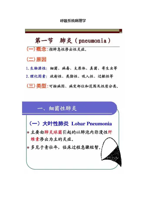
呼吸系统病理学呼吸系统疾病第一节慢性阻塞性肺病一、慢性支气管炎(chronic bronchitis)是指气管、支气管粘膜及其周围组织的慢性非特异性炎症。
临床上以反复咳嗽、咳痰或伴有喘息症状为特征,且症状每年至少持续3个月,连续两年以上。
(一)病因和发病机制:感冒;寒冷气候;病毒感染和继发性细菌感染;最常见的病毒是鼻病毒、腺病毒和呼吸道合胞病毒,细菌最常见的是肺炎球菌、肺炎克雷伯杆菌、流感嗜血杆菌、奈瑟球菌等。
吸烟;长期接触工业粉尘、大气污染和过敏因素;内在因素:机体抵抗力降低,呼吸系统防御功能受损、神经内分泌功能失调。
(二)病理变化:各级支气管均可受累。
反复的损伤和修复。
1、粘膜上皮病变纤毛倒伏、脱失。
上皮细胞变性、坏死脱落,杯状细胞增多,并可发生鳞状上皮化生。
2、粘液腺肥大、增生,分泌亢进,浆液腺发生粘液化。
3、管壁充血,淋巴细胞、浆细胞浸润;4、管壁平滑肌束断裂、萎缩,软骨变性、萎缩,钙化或骨化。
(三)临床病理联系:咳嗽、咳痰、喘息。
痰一般呈白色粘液泡沫状。
二、肺气肿:肺气肿(pulmonary emphysema)是指呼吸细支气管以远的末梢肺组织(包括呼吸性支气管、肺泡管、肺泡囊、肺泡)因残气量增多而呈持久性扩张,并伴有肺泡间隔破坏,以致肺组织弹性减弱,容积增大的一种病理状态。
(一)病因和发病机制:1、阻塞性通气障碍慢性支气管炎等细支气管炎症--造成管壁增厚,管腔狭窄,阻塞或塌陷。
2、老年性肺组织发生退行性变,弹性回缩力降低而引起3、抗胰蛋白酶缺乏导致弹性蛋白酶增多、活性增高—降解肺组织中的弹性蛋白、胶原蛋白和蛋白多糖,导致肺的组织结构受损。
先天性常染色体隐性遗传病,发病年龄早,有家族史,病程短,多为全腺泡型肺气肿。
(二)类型及其病变特点:1、肺泡性肺气肿由于常合并有小气道阻塞性通气障碍,故又有慢性阻塞性肺气肿之称。
(1)腺泡中央型肺气肿肺腺泡中央区的呼吸性细支气管囊状扩张,肺泡管、肺泡囊变化不明显。
病理学呼吸系统疾病一、判断题1 .肺小叶内终末细支气管及其远端所属的肺组织称为肺腺泡。
[判断题] *对错(正确答案)1 .肺小叶内终末细支气管及其远端所属的肺组织称为肺腺泡。
[判断题] *对错(正确答案)2 .吸烟是引起慢性支气管炎、肺气肿和肺癌的主要原因之一。
[判断题] *对(正确答案)错3慢性支气管炎病人咳嗽、咳痰的病变基础主要是支气管壁粘液腺增生、肥大和浆液腺粘液化生。
[判断题] *对(正确答案)错4.阻塞性肺气肿的发生主要是由于小气道的炎症和阻塞性通气障碍,以及肺间隔的破坏所致。
[判断题] *对(正确答案)错5.代偿性肺气肿和老年性肺气肿不是真性肺气肿。
[判断题] *错6 .迟发性支气管哮喘的发作主要与T淋巴细胞和嗜酸性粒细胞有关。
[判断题] *对错(正确答案)7.支气管扩张症是指肺内支气管管腔永久性扩张伴管壁纤维性增厚的一种慢性化脓性疾病。
[判断题] *对(正确答案)错8慢性支气管炎与支气管扩张症的病变差别之一是后者支气管粘膜上皮不发生鳞状化生。
[判断题] *对错(正确答案)9.大叶性肺炎的病变主要是肺泡的纤维素性炎,因而常合并肺间质纤维化。
[判断题] *对错(正确答案)10 .大叶性肺炎充血水肿期和红色肝样变期,在肺泡内渗出液中能检出多量肺炎球菌 [判断题] *对(正确答案)错11 .在大叶性肺炎灰色肝样变期,肺实变程度最重,故病人缺氧的症状也最明显。
[判断题] *错12 .当肺内癌组织弥漫浸润使肺实变、肉眼观呈肉质样改变时称为肺肉质变。
[判断题] *对错(正确答案)13 .小叶性肺炎是以细支气管为中心、肺小叶为单位的急性化脓性炎症,常只累及一叶肺组织。
[判断题] *对错(正确答案)14 .严重的小叶性肺炎,病灶可相互融合,形成融合性小叶性肺炎,若不抓紧治疗,有可能发展为大叶性肺炎。
[判断题] *对错(正确答案)15.小叶性肺炎好发于儿童和年老体弱者,且常常是某些疾病的并发症。
1、慢性支气管炎患者通气功能障碍得病理基础就是A、粘液腺肥大、增生B、上皮纤毛倒伏、脱落C、软骨变性萎缩D、小气道阻塞E、管壁平滑肌萎缩;2、引起支气管哮喘得主要原因就是A、支气管粘膜内杯状细胞增多B、支气管粘膜水肿C、支气管壁平滑肌痉挛D、支气管腔内粘液增多E、支气管管壁炎细胞浸润;3、全腺泡型肺气肿得主要原因就是A、小气道阻塞B、支气管壁炎细胞浸润C、肺组织炎细胞浸润D、先天性?1-抗胰蛋白酶缺乏E、支气管壁扩张;4、下列疾病中,最常引起肺心病者就是A、肺动脉栓塞B、胸廓畸形C、矽肺D、慢性肺结核E、肺气肿;5、不符合支气管扩张特点得就是A、多发生于叶及段等大支气管B、支气管壁得炎症损伤就是主要发病基础C、肺脓肿为其常见合并症D、可导致肺心病E、临床表现咳大量脓痰及反复咯血;6、不符合小叶性肺炎得就是A、主要由病毒引起B、小叶与小叶融合性病变C、病变以肺得背部与下部较重D、可导致心力衰竭E、可引起肺脓肿;7、大叶性肺炎得合并症有A、肺肉质变B、感染性休克C、纤维素性胸膜炎D、肺脓肿E、以上都就是; 8、病毒性肺炎得确诊主要依据就是A、X线检查B、血清学检查C、病毒与病毒抗原得检查D、病毒感染流行状况E、儿童与老年人;9、确诊支原体肺炎得可靠依据就是A、X线检查B、血清学检查C、病原学检查D、支原体感染流行状况E、排除细菌性肺炎得可能;10、不符合绿脓杆菌肺炎特点得就是A、易引起败血症B、多呈小叶性病变C、多见于社区性肺炎D、多发生于严重烧伤、白血病、化学治疗患者E、多引起肺脓肿;11、符合矽结节得描述就是A、早期就是纤维结节B、晚期就是细胞结节C、其发生与T淋巴细胞关系密切D、具有病理诊断意义E、不含免疫球蛋白;12、不符合农民肺得描述就是A、由于吸入高温放线菌引起得过敏性肺泡炎B、肺内出现肉芽肿病变C、类上皮细胞内可见放线菌孢子D、肺间质纤维组织增生E、属Ⅰ型超敏反应性疾病;13、最常见得鼻咽癌得组织学类型就是A、未分化癌B、泡状核细胞癌C、腺泡状腺癌D、低分化鳞状细胞癌E、高分化鳞状细胞癌;14、小叶性肺炎得病变性质主要就是A、卡她性炎B、纤维素性炎C、肉芽肿性炎D、化脓性炎E、浆液性炎;15、不符合肺腺癌得描述就是A、多为周围型B、常有吸烟史C、可伴有粘液分泌D、常有肺门淋巴结转移E、常有乳头形成;16、肺癌得主要组织来源就是A、支气管上皮细胞B、Clara细胞C、Kultschitzky细胞D、Ⅰ型肺泡上皮细胞E、Ⅱ性肺泡上皮细胞;17、患者突起畏寒高热、胸痛、咳铁锈色痰,最有可能就是A、慢性支气管炎B、大叶性肺炎C、肺结核D、肺心病E、支气管扩张症;18、下列哪项不符合小叶性肺炎A、可由多种细菌感染引起B、可导致支气管扩张C、病灶直径多在1cm左右D、病灶融合发展为肺肉质变E、支气管与肺组织遭破坏;19、男性,50岁,近一月来感到胸部胀闷,近3天伴有气促、心慌。

第一节肺炎〔Pneumonia〕1、概念:肺部的血管系统对各种致炎因子产生防御反响所表现出来的急性渗出性炎症。
多指终末气道、泡和肺间质的炎症。
2、分类:●按病因:细菌性、病毒性、支原体性●按范围:大叶性、小叶性、间质性●按性质:纤维素性、化脓性、出血性肉、芽肿性、浆液性、干酪性一、大叶性肺炎〔lobar pneumonia〕●主要是由肺炎链球菌感染引起,病变起始于肺泡,并迅速扩展至整个或多个大叶的肺的急性弥漫性纤维素性渗出性炎。
●主要表现:起病急、寒颤、高热、胸痛、铁锈色痰、血白细胞上升、咳嗽、呼吸困难、肺实变体征,青壮年〔男性〕多见。
〔一〕病因和发病机制:1、病因:90~95%由肺炎链球菌〔或肺炎杆菌金葡溶血链流感嗜血〕引起,机体抵抗力降低时发病。
2、发病机理:肺炎球菌有荚膜,对组织有抗吞噬作用。
机体对肺炎球菌有过敏现象。
细菌通过变态反响,使肺泡壁毛细血管通透性增加,导致毛细血管扩张,大量浆液、纤维蛋白原渗出引起纤维素性渗出性炎。
病变开展迅速,炎症经肺泡孔、支气管累及肺段或整个肺叶。
而局部不一定有肺炎球菌的出现。
〔二〕病理改变及临床病理联系:1、病理改变:肺泡壁毛细血管的扩张和闭塞;肺泡腔内渗出物内容的改变。
2、分期及特点:〔1〕充血水肿期〔congestion〕:肺泡壁毛细血管扩张充血,肺泡腔内有大量浆液性渗出物〔混有少数红细胞、中性粒细胞和巨噬细胞,并含有大量细菌〕。
肉眼观:病变肺叶充血、水肿、呈暗红色。
临床病理联系:细菌〔+〕:毒血症〔2〕红色肝样变期〔red hepatization〕:肺泡壁毛细血管扩张充血,肺泡腔内渗出物有大量红细胞及纤维素。
肉眼观:病变肺叶呈暗红色,切面成颗粒状,质地变实,类似于肝。
肺实变〔consolidation of lung〕:任何原因致肺泡腔内积聚浆液、纤维蛋白和细胞成分等,使肺泡含气量减少、肺质地致密化的一种病变。
临床病理联系:①细菌〔+〕:毒血症;②胸膜渗出:胸痛、摩擦音;③红细胞:铁锈色痰;④无效灌注:缺氧〔3〕灰色肝〔样〕变期〔gray hepatization〕:肺泡壁毛细血管闭塞,肺泡腔内渗出物以纤维素为主,还有大量中性粒细胞。

三基练习-病理学-第10章呼吸系统疾病+答案+完整解析1、下列关于大叶性肺炎的描述,错误的是:A、由细菌感染引起B、病变多从肺泡开始C、炎症不破坏肺泡壁结构D、患者常有胸痛及咳铁锈色痰E、中性粒细胞渗出为主的炎症解析:大叶性肺炎是以纤维素渗出为主的炎症。
正确答案:E2、大叶性肺炎的并发症不包括()A、蜂窝肺B、脓气胸C、纤维素性胸膜炎D、肺肉质变E、败血症和脓毒败血症解析:大叶性肺炎的并发症:①肺肉质变;②肺脓肿或脓胸;③胸膜肥厚或粘连:由纤维素性胸膜炎不能被完全溶解吸收引起;④败血症或脓毒败血症;⑤感染性休克。
正确答案:A3、中性粒细胞渗出过少的大叶性肺炎易发生的并发症是()A、感染性休克B、脓胸C、纤维素性胸膜炎D、肺肉质变E、败血症解析:大叶性肺炎病灶中的中性粒细胞渗出过少,渗出物不能完全溶解而发生机化,形成肺肉质变。
正确答案:D4、渗出液中检出肺炎球菌,提示大叶性肺炎病变处于()A、充血水肿期B、红色肝样变期和灰色肝样变期C、灰色肝样变期D、溶解消散期E、充血水肿期和红色肝样变期解析:灰色肝样变期时,渗出物中有大量中性粒细胞且特异性抗体已形成,可吞噬杀灭致病菌,故导致病菌不易检出;溶解消散期,肺内的炎症已经消散溶解,更不易检出致病菌。
正确答案:E5、患儿,男,2岁。
近几日出现发热、咳嗽,伴黄色黏液脓痰,胸透见两肺下叶散在边缘模糊的斑片状阴影。
其最可能的病变是A、干酪样肺炎B、大叶性肺炎C、小叶性肺炎D、间质性肺炎E、转移性肿瘤正确答案:C6、下列不属于小叶性肺炎的是()A、手术后肺炎B、吸入性肺炎C、麻疹后肺炎D、坠积性肺炎E、巨细胞肺炎解析:麻疹性肺炎时出现的巨细胞较多,又称巨细胞肺炎,为病毒性肺炎。
正确答案:E7、下列病理改变符合病毒性肺炎的是A、常有肺泡结构的破坏B、肺泡间隔水肿,炎细胞浸润C、肺泡内常有大量纤维素渗出D、局部区域中性粒细胞浸润E、一般不检出病毒包涵体解析:病毒性肺炎镜下表现主要为肺间质的炎症,而非肺泡结构的破坏。
病理学之呼吸系统疾病试题及答案一、选择题单选题1.下列哪项不符合小叶性肺炎A.多由致病力弱的肺炎球菌引起B.好发于老人、儿童、久病卧床者C.以细支气管为中心的纤维素性炎症D.常作为其它疾病的并发症出现E.病灶可互相融合2.鼻咽癌的常见组织学类型A.高分化鳞癌B.泡状核细胞癌C.未分化癌D.腺癌E.低分化鳞癌3.大叶性肺炎的病变性质是:A.纤维素性炎B.变态反应性炎C.化脓性炎D.浆液性炎E.出血性炎4.鼻咽癌常发生在A.鼻咽后部B.鼻咽顶部C.鼻咽侧壁D.鼻咽前壁E.鼻咽底部5.肺源性心脏病最常见的原因是A.支气管哮喘B.支气管扩张C.慢性支气管炎D.肺结核病E.矽肺6.小叶性肺炎的病变范围A.以呼吸性细支气管为中心B.以终未细支管为中心C.以细支气管为中心D.以支气管为中心E.以肺泡管为中心7.二氧化硅尘致病力最强的是A.<5微米B.>5微米C.<3微米D.1-2微米E.3-4微米8.鼻咽泡状核细胞癌哪一项是错的?A.胞体大,胞浆丰富B.细胞多边形境界清楚C.核大、染色质小,空泡状D.核仁肥大、可1-2个E.癌细胞间有淋巴细胞浸润9.某男,60岁、胸痛、咳嗽、咯血痰两个月,胸片见右上肺周边一直径为5cm结节状阴影,边缘毛刺状。
应首先考虑:A.肺结核球B.周围型肺癌C.团块状矽结节D.肺脓肿E.肺肉质变10.硅肺的并发症有A.肺脓肿B.肺结核C.肺癌D.肺炎E.肺结核+肺心病11.下列低分化鼻咽癌的临床病理特征中,哪一项不正确?A.原发灶往往很小B.早期可有同侧颈淋巴结转移C.对放射治疗不敏感D.以低分化鳞癌最常见E.易侵犯颅底及颅神经12.肺心病发病的主要环节是:A.慢性支气管炎B.慢性阻塞性肺气肿C.肺纤维化D.肺血管床减少E.肺循环阻力增加和肺动脉高压13.大叶性肺炎的肉质变是由于A.中性白细胞渗出过多B.中性白细胞渗出过少C.纤维蛋白原渗出过多D.红细胞漏出过多E.红细胞漏出过少14.大叶性肺炎的病变特点.下列哪项除外A.病变中可有大量细菌B.较多的红细胞漏出C.大量纤维素渗出D.常有肉质变E.大量中性白细胞渗出15.小叶性肺炎的病变性质多为:A.出血性纤维素性炎症B.卡他性炎症C.增生性炎症D.化脓性炎症E.变质性炎症16.肺癌最常见的组织学类型是:A.腺样囊性癌B.巨细胞癌C.鳞状细胞癌D.腺癌E.未分化癌多选题1.慢性支气管炎可导致A.支气管扩张症B.支气管管腔狭窄C.肺气肿D.支气管性哮喘2.鼻咽癌的特点是A.早期就可发现鼻咽部明显肿块B.以低分化鳞癌最为多见C.未分化癌大多来自粘膜上皮的嗜银细胞D.往往早期发生淋巴道转移3.肺源性心脏病是由下列疾病所引起A.间质性肺炎所致的间质纤维化B.反复发作的慢性细支气管炎及细支气管周围炎C.间质性肺气肿D.支气管扩张症4.硅肺对人的危害在于A.引起肺心病B.并发肺结核C.引起肺纤维化影响气体交换D.晚期常发肺癌5.哪些是慢性支气管炎的典型病变A.粘膜腺体肥大增生B.平滑肌肥大C.杯状细胞增生D.支气管壁内嗜酸性白细胞浸润参考答案:单选题:1、C E A B C 6、C D B B E 11、C E B D D 16、C多选题:1、ABC2、BD3、ABD4、ABC5、AC二、是非题1. 燕麦细胞癌属于APUD瘤,其组织学来源为支气管粘膜上皮细胞。