外泌体科学研究策略与方法
- 格式:pptx
- 大小:8.65 MB
- 文档页数:38

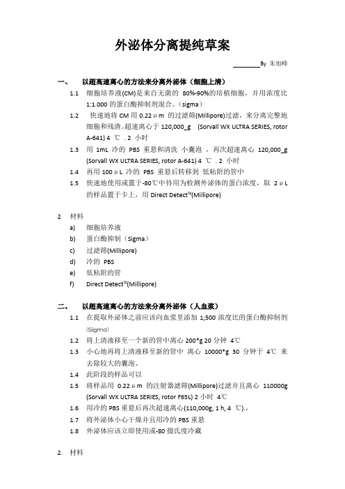
外泌体分离提纯草案By 朱旭峰一、以超高速离心的方法来分离外泌体(细胞上清)1.1细胞培养液(CM)是来自无菌的80%-90%的培植细胞,并用浓度比1:1.000的蛋白酶抑制剂混合。
(sigma)1.2快速地将CM用0.22μm 的过滤筛(Millipore)过滤,来分离完整地细胞和残渣。
超速离心于120,000_g (Sorvall WX ULTRA SERIES, rotorA-641) 4 ℃. 2 小时1.3用1mL 冷的PBS 重悬和清洗小囊泡,再次超速离心120,000_g(Sorvall WX ULTRA SERIES, rotor A-641) 4 ℃. 2 小时1.4再用100μL 冷的PBS 重悬后转移到低粘附的管中1.5快速地使用或置于-80℃中待用为检测外泌体的蛋白浓度,取2μL的样品置于卡上,用Direct Detect™(Millipore)2材料a)细胞培养液b)蛋白酶抑制(Sigma)c)过滤筛(Millipore)d)冷的PBSe)低粘附的管f)Direct Detect™(Millipore)二、以超高速离心的方法来分离外泌体(人血浆)1.1在提取外泌体之前应该向血浆里添加1;500浓度比的蛋白酶抑制剂(Sigma)1.2将上清液移至一个新的管中离心200*g 20分钟4℃1.3小心地再将上清液移至新的管中离心10000*g 30分钟于4℃来去除较大的囊泡、1.4此阶段的样品可以1.5将样品用0.22μm的注射器滤筛(Millipore)过滤并且离心110000g(Sorvall WX ULTRA SERIES, rotor F65L) 2小时4℃1.6用冷的PBS重悬后再次超速离心(110,000g, 1 h, 4 ℃).,1.7将外泌体小心干燥并且用冷的PBS重悬1.8外泌体应该立即使用或-80摄氏度冷藏2.材料a)蛋白酶抑制(Sigma)b)过滤筛(Millipore)c)冷的PBSd)低粘附的管e)Direct Detect™(Millipore)三、ExoQuick TM化学沉淀法1.1ExoQuick TM的使用要按照厂家提供的说明书来实行1.2该试剂盒可以简单地提取CM,人血浆,和血清里的外泌体,只需用倒相管按照所指示的量来加入ExoQuick TM试剂盒里的溶液1.3溶液孵化过夜,在4℃温和的摇晃1.4在流式细胞仪缓冲液中洗涤1.5试剂盒里的带荧光的免疫磁珠已经被绑定,用流式细胞仪鉴定即可(BD Biosciences) 配合使用CellQuest 的软件。
外泌体作为新型治疗手段的研究随着科技的不断进步,越来越多的新型治疗手段被研究出来,其中外泌体作为一种新兴的治疗手段,受到了科学家们的广泛关注。
外泌体是一种由细胞分泌出来的纳米级贝壳结构的小囊泡,内部含有具有生物活性的基因信息和蛋白质等分子物质。
近年来,外泌体在治疗癌症、心血管疾病、神经退行性疾病等方面的应用研究也得到了广泛的关注。
外泌体作为新型的治疗手段,有着许多独特的特性。
首先,作为一种纳米级的小囊泡,外泌体能够穿透生物膜,有效地将其内部的物质传递到细胞内。
其次,外泌体内部含有多种生物活性物质,如蛋白质、mRNA、miRNA、DNA等,这些生物活性分子可以被利用来转化细胞功能或修饰基因表达。
最后,由于外泌体来源于自体细胞,也不涉及使用外来材料或免疫反应问题,因此具有良好的安全性和免疫学适应性。
在治疗癌症方面,外泌体已被证实具有抗癌活性。
研究人员发现,肿瘤细胞和其他细胞相比,分泌的外泌体数量更多,且含有更多的生物活性物质和生长因子,这些物质具有促进肿瘤细胞增殖和侵袭的作用。
因此,通过拦截肿瘤细胞分泌的外泌体或直接给予患者来源于其他细胞的外泌体,可以达到治疗癌症的效果。
例如,研究人员发现通过注射来源于骨髓间充质干细胞的外泌体,可以抑制恶性胶质瘤的生长,并提高患者的存活率。
在治疗心血管疾病方面,外泌体也具有广泛的应用前景。
心肌缺血后心肌细胞将大量释放外泌体,其中包含补体激活物和血小板激活因子等生物活性物质。
通过注射来源于心肌细胞外泌体的治疗方法可以提高心肌细胞的功能和心脏的整体状态。
同时,研究人员也发现,通过从骨髓间充质干细胞收集分离出的外泌体可以改善心血管纤维化、促进血管新生和心肌再生,从而有效改善心血管的健康状况。
在神经退行性疾病的治疗中,外泌体也有着潜在的作用。
神经元为了维持生命所需要的蛋白质能够从周围的胶质细胞中获得,但由于神经元本身的限制,导致神经毒性事件的发生,这也增加了神经退行性疾病的发生率。

外泌体的研究进展一、本文概述随着生物医学研究的深入,细胞间的信息交流机制逐渐成为研究的热点。
作为细胞间交流的重要载体,外泌体(Exosomes)的研究在近年来取得了显著的进展。
本文旨在综述外泌体的基本特征、生物学功能及其在疾病诊断和治疗中的应用,同时探讨当前面临的挑战和未来的发展趋势。
我们将简要介绍外泌体的定义、结构特点以及产生机制,帮助读者理解其作为细胞间信息传递的重要角色。
接着,我们将重点讨论外泌体在肿瘤、心血管疾病、神经退行性疾病等医学领域的研究进展,包括外泌体在疾病发生发展中的作用机制,以及作为疾病标志物的潜力。
我们还将关注外泌体在疾病诊断和治疗中的应用前景,如作为药物递送载体、肿瘤疫苗以及生物标志物等方面的研究。
我们将对当前外泌体研究面临的挑战和未来的发展方向进行深入探讨,以期为推动外泌体在生物医学领域的应用提供有益的参考。
二、外泌体的结构与功能外泌体是一种由细胞主动分泌的,直径约为30-150纳米的膜性囊泡,普遍存在于各种体液中,包括血液、尿液、乳汁、脑脊液和细胞培养基等。
这些囊泡在细胞间的物质传递、信息交流以及免疫反应等方面发挥着重要作用。
近年来,随着外泌体研究的深入,其独特的结构和功能逐渐被人们所揭示。
结构上,外泌体由磷脂双分子层膜包裹着内部的水溶液组成,其膜上嵌有多种蛋白质,包括四跨膜蛋白、热休克蛋白、整合素等。
这些蛋白质不仅参与外泌体的形成和分泌过程,还负责将外泌体与靶细胞进行特异性结合,实现精准的物质传递。
外泌体的膜上还含有丰富的糖类和脂质,这些成分对于外泌体的稳定性和功能也至关重要。
功能上,外泌体具有多种生物学活性。
外泌体可以传递信息分子,如mRNA、miRNA、蛋白质等,这些分子在细胞间的信息交流过程中发挥着关键作用。
外泌体可以参与免疫反应,通过传递抗原或免疫调节分子,影响免疫细胞的活性和功能。
外泌体还具有促进血管生成、抑制肿瘤生长等多种生物学活性。
值得一提的是,外泌体的功能与其来源细胞密切相关。
外泌体在生理与病理过程中的作用及研究进展外泌体是指一种细胞分泌的小囊泡,直径约为30-100纳米。
它们可以携带细胞膜、细胞器质膜和多种生物活性分子,比如蛋白质、miRNA和毒素,通过与其他细胞交互影响它们的行为。
在最近的几年,外泌体的发现引起了广泛的兴趣,因为它们被认为在生理和病理过程中扮演了关键的角色。
外泌体的形成和释放外泌体从各种类型的细胞中分泌,包括血细胞、肝细胞、上皮细胞和神经细胞等。
它们可以通过三种主要的机制来形成和释放:直接分泌、外化和内化。
直接分泌方式是外泌体在细胞膜上形成,然后释放到细胞外。
外化是指外泌体与细胞膜融合从而形成它们的过程。
内化是指细胞通过内吞作用摄取外泌体。
外泌体的功能外泌体具有多种功能,它们可以在生理和病理过程中发挥不同的作用,具体如下:1. 细胞通信:外泌体可以通过携带生物活性分子来影响接收者细胞的行为,从而参与细胞通信。
例如,蛋白质可以调节受体表达、代谢和细胞增殖等。
2. 细胞凋亡:外泌体可以参与调控细胞凋亡。
一个细胞可以释放外泌体包含有调节细胞凋亡的分子,例如Caspace9,就可以影响周围细胞的凋亡和存活。
3. 免疫调节:外泌体作为一类重要的信息介质扮演着免疫细胞之间互相沟通交流的桥梁。
外泌体中的mRNA和miRNA可以通过调节靶基因的表达,影响细胞的生物学特性和免疫反应。
外泌体在疾病发展中的作用1. 肿瘤:外泌体在肿瘤生长和转移中发挥重要作用。
外泌体在内部远程传递肿瘤信息,可以调节肿瘤细胞的生长、转移和侵染,并参与肿瘤微环境的建立。
研究发现,肿瘤患者血浆中的外泌体数量和质量都发生了改变,这提示外泌体对肿瘤病理生理过程具有极大的影响。
2. 神经性疾病:外泌体也可以在神经细胞之间传递信息。
研究显示在神经元发展中,外泌体可以调节同种或异种神经元的扩散。
这些影响也被发现与神经退行性疾病如阿兹海默症,帕金森氏病,运动神经元疾病如渐冻人有关。
3. 心血管疾病:外泌体在心血管系统中的病理过程中的作用也在不断被探究。
外泌体的研究进展外泌体是细胞分泌的一种重要物质,由细胞释放到细胞外环境中,参与多种生物学过程。
近年来,随着对外泌体研究的深入,越来越多的学者开始其在医学、生物和材料等领域的应用价值。
本文将对外泌体的研究现状、方法、成果和应用前景进行全面探讨。
一、外泌体的研究现状外泌体是由细胞分泌的一种小泡,直径约30-100纳米,包含多种生物活性物质,如蛋白质、核酸和脂质等。
近年来,随着对外泌体的深入研究,发现其参与了细胞间通信、物质运输等多种生物学过程。
同时,外泌体在疾病诊断、药物研发和组织工程等方面也具有广泛的应用前景。
二、外泌体的研究方法1、实验设计:外泌体的研究涉及细胞生物学、分子生物学、生物化学、医学等多个学科,因此需要进行多学科交叉的实验设计。
2、样本处理:外泌体的提取和纯化是研究外泌体的关键步骤,常用的方法包括超速离心法、免疫磁珠法、聚合物沉淀法等。
3、基因表达分析:采用基因表达谱技术对外泌体中的核酸进行分析,以揭示其基因表达特征。
4、蛋白质组学:运用蛋白质组学技术对外泌体中的蛋白质进行分析,以鉴定其蛋白质种类和功能。
三、外泌体的研究成果1、肿瘤治疗:研究发现,肿瘤细胞释放的外泌体可以传递信号给正常细胞,诱导其恶性转化。
因此,通过研究外泌体在肿瘤发生发展中的作用,有望为肿瘤治疗提供新思路。
2、药物传输:外泌体具有保护和输送物质的特性,可被用作药物载体。
通过加载抗癌药物或其他生物活性物质,外泌体可以实现药物的定向传输,提高疗效并降低副作用。
3、组织工程:外泌体具有调节细胞生长、分化和迁移的能力,因此在组织工程中具有潜在应用价值。
例如,将特定外泌体加载到生物材料中,可以促进组织再生和修复。
4、环境监测:外泌体在细胞间通信和物质运输中发挥重要作用,也参与了环境因素的响应和适应。
通过研究外泌体在环境监测中的作用,可以为环境污染的监测和治理提供新手段。
四、外泌体的应用前景1、生物治疗手段:通过调节外泌体释放的生物活性物质,有望开发新的生物治疗手段,如上文提到的肿瘤治疗和药物传输等。

人骨髓来源间充质干细胞分泌外泌体特性研究一、本文概述Overview of this article随着生物医学领域的快速发展,干细胞及其分泌产物的研究已成为当前生命科学领域的热点之一。
人骨髓来源间充质干细胞(hMSCs)作为一种具有多向分化潜能的成体干细胞,其在组织工程、再生医学以及疾病治疗等领域的应用潜力备受关注。
近年来,随着对外泌体(Exosomes)研究的深入,人们发现hMSCs分泌的外泌体在细胞间的信息传递、免疫调节以及组织修复等方面发挥着重要作用。
因此,本文旨在深入研究人骨髓来源间充质干细胞分泌外泌体的特性,以期为hMSCs在医学领域的临床应用提供新的思路和方法。
With the rapid development of the biomedical field, research on stem cells and their secreted products has become one of the hotspots in the current field of life sciences. Human bone marrow-derived mesenchymal stem cells (hMSCs), as an adult stem cell with multi-directional differentiation potential, have attracted much attention for their potential applications in tissue engineering, regenerative medicine, and diseasetreatment. In recent years, with the deepening of research on exosomes, it has been found that exosomes secreted by hMSCs play important roles in intercellular information transmission, immune regulation, and tissue repair. Therefore, this article aims to investigate in depth the characteristics of human bone marrow-derived mesenchymal stem cells secreting exosomes, in order to provide new ideas and methods for the clinical application of hMSCs in the medical field.本文将首先介绍hMSCs的基本生物学特性及其在组织工程和再生医学中的应用。
外泌体科学研究策略与方法外泌体(extracellular vesicles)是一类由细胞分泌的小型膜囊泡,具有丰富的生物大分子(如蛋白质、核酸和代谢产物等)和受体分子,能够通过体液循环进入其他细胞并调控其状态。
外泌体作为一种新兴的细胞间通讯方式,已经在生物学和医学研究中受到广泛关注。
为了深入研究外泌体的功能和应用价值,科学家们采用了一系列研究策略和方法。
首先,外泌体的纯化和鉴定是外泌体研究的基础。
目前常用的外泌体纯化方法包括超速离心、滤膜浓缩、亲和析取和尺寸排除等。
其中,超速离心是最常用的方法,通过连续离心将外泌体从细胞碎片、蛋白质聚集体和其他细胞分泌物中分离出来。
纯化后的外泌体可以通过电镜观察其形态和大小,并通过蛋白质和核酸的检测确认其特征标志物。
其次,外泌体内的生物大分子是外泌体功能的关键。
外泌体中的蛋白质、核酸和代谢产物等分子可以通过质谱分析、RNA测序、代谢组学等方法进行全面的分析。
通过对外泌体中的蛋白质组学的研究,可以鉴定外泌体特有的标志物,并推测其功能和信号通路。
通过外泌体中的RNA测序,可以发现外泌体携带的mRNA和miRNA,了解其在细胞间通讯中的作用。
通过代谢组学的研究,可以分析外泌体携带的代谢产物,揭示其在代谢调控中的作用。
第三,外泌体功能研究是外泌体研究的重点之一、外泌体功能研究主要包括外泌体的分泌机制、目标细胞的识别和摄取、信号传递和功能调控等方面。
通过抑制外泌体分泌的相关因子,可以阻断外泌体的释放,从而探究外泌体的分泌机制。
通过标记外泌体表面的受体和底物,可以观察其在目标细胞上的相互作用和摄取情况。
通过干预外泌体释放和摄取的相关蛋白质和信号通路,可以揭示外泌体在细胞间通讯中的功能和调控机制。
最后,外泌体在生理和病理过程中的应用是外泌体研究的重要方向之一、外泌体作为一种重要的细胞间信号载体,可以通过改变外泌体的组成和功能来调控细胞的生理状态。
通过人工制备外泌体,可以将特定的蛋白质、核酸和药物等载体到外泌体中,改变外泌体的功能并进一步实现治疗效果。
外泌体研究思路解析外泌体(extracellular vesicles,EVs)是一类由细胞分泌的膜囊泡,其直径一般在30-150纳米之间。
近年来,外泌体的研究受到了广泛关注,并且被证明在各种生物学过程中起着重要的作用,包括细胞间通信、免疫调节、细胞增殖和转化等。
在这篇文章中,我将介绍外泌体的研究思路,并阐述其在生物医学研究中的应用前景。
其次,研究者可以通过外泌体的分离和纯化来进一步探索其功能和应用。
目前,常用的外泌体分离方法包括差速离心、滤膜分离和亲和纯化等。
这些方法能够选择性地分离不同类型的外泌体,并提取其纯净的样品,以便于后续的功能分析和应用研究。
从纯化的外泌体中,研究者可以通过电子显微镜、免疫荧光染色和质谱分析等方法来进行外泌体的表征和定量。
在功能研究方面,研究者可以通过体外细胞培养等方式,研究外泌体对目标细胞的调控作用。
例如,外泌体中的特定蛋白质和RNA分子可以通过细胞内信号通路传递,从而调节目标细胞的增殖、分化和凋亡等生物学过程。
同时,通过外泌体的功能分析,研究者还可以了解外泌体在疾病中的作用,以及用于疾病诊断和治疗的潜在应用。
最后,外泌体的应用前景广阔,可以用于生物医学研究的多个领域。
首先,外泌体作为细胞间的信息传递媒介,可以用于药物递送和基因治疗等领域。
其次,外泌体中的蛋白质和RNA等分子可以作为生物标志物,用于疾病的诊断和预后评估。
此外,外泌体还可以用于生物治疗的制备,例如利用外泌体激活免疫系统来对抗癌症等疾病。
综上所述,外泌体的研究思路包括了对其生物学特性和生物合成机制的探究、外泌体的分离和纯化、外泌体的功能分析以及外泌体的应用前景探究。
通过深入研究外泌体,我们可以更好地理解其在生物学过程中的作用,为生物医学研究提供新的思路和方法。

外泌体提取方法及鉴定分析研究进展外泌体是细胞分泌的一种微小膜泡,包含了多种生物活性分子,如蛋白质、核酸和脂质等。
近年来,随着生物技术的不断发展,外泌体提取方法和鉴定分析研究取得了显著的进展。
本文将对外泌体提取方法及鉴定分析研究进展进行简要综述。
外泌体是由细胞分泌的一种直径约30-100nm的膜泡,由细胞内吞摄取并加工处理形成。
外泌体在细胞间通讯、物质运输以及疾病诊断等方面具有重要作用。
化学法是一种常用的外泌体提取方法,主要采用离心和沉淀相结合的方法。
首先将细胞培养液进行高速离心,去除细胞和较大的膜泡,然后加入沉淀剂沉淀获得外泌体。
该方法的优点是操作简单、提取量较大,适合大量样本的处理。
但是,该方法纯度较低,可能会影响后续的鉴定和分析。
生物法是一种利用细胞表面标志物进行免疫吸附的外泌体提取方法。
将外泌体进行免疫吸附,再通过洗脱得到纯度较高的外泌体。
该方法的优点是纯度高、对细胞损伤小,适用于珍贵样本的提取。
但是,该方法操作复杂,提取量较小,需要大量的抗体。
基因工程法是一种利用细胞表达特定基因的外泌体提取方法。
将目的基因转染到细胞中,通过表达特定膜蛋白,诱导细胞分泌出外泌体,再对其进行纯化和提取。
该方法的优点是纯度高、提取量较大,适用于具有特定功能的外泌体提取。
但是,该方法需要特定的基因工程技术和细胞系,操作较为复杂。
蛋白质组学在外泌体研究中的应用不断深入。
通过蛋白质组学技术,可以鉴定外泌体中的蛋白质种类、相对丰度以及修饰情况等。
通过比较不同条件下外泌体蛋白质组学的差异,可以研究外泌体的生物学特征和功能。
随着二代测序技术的不断发展,基因序列分析在外泌体研究中也得到广泛应用。
通过对外泌体中的核酸进行测序,可以鉴定其中的基因序列和表达水平。
通过比较不同条件下外泌体基因序列的差异,可以研究外泌体在细胞间通讯和疾病诊断等方面的作用。
转录组测序转录组测序可以分析外泌体中的mRNA、lncRNA和miRNA等转录本。

植物外泌体途径的分子机制研究随着科技的不断发展,人们对于植物的了解也越来越深刻。
近年来,越来越多的研究发现,植物细胞外泌体作为最重要的信息素传递途径之一,在植物的生态和生理过程中扮演着重要的角色。
植物外泌体途径涉及到许多与生物学和生物化学相关的重要机制,是目前生命科学领域研究的热点和难点之一。
本文将从植物外泌体的特征、来源以及分子机制等方面进行探究,以期对植物外泌体途径的研究起到一定的启发和帮助。
一、植物外泌体的特征植物外泌体是指一种细胞分泌物,主要由植物细胞质浆中移动到植物细胞表面,形成特定的膜结构,包含了丰富的生物信息素分子和由细胞壁成分构成的蛋白质。
植物外泌体的大小一般在30-100纳米之间,通常分为四种类型:基质分泌体、粘质分泌体、普通壁分泌体和小囊泡分泌体。
通常来说,植物外泌体的表面会覆盖有糖蛋白、脂质、磷脂酰肌醇等成分,这些成分能够与其他细胞或分子发生互相作用,将信号、蛋白质或其他生物分子等从一些细胞分泌到另一些细胞内。
二、植物外泌体的来源植物外泌体的生成思维复杂和动态的过程。
植物细胞通过胞吐作用将所需的蛋白质、RNA、食物等分子从胞内传输到胞外,形成特定的细胞膜结构,进而形成外泌体。
这些膜结构包含了丰富的复杂蛋白质和其他有机物,从而形成涉及到许多分子机制的复杂系统。
三、植物外泌体途径的分子机制许多研究表明,植物外泌体途径的分子机制涉及到许多重要的分子和信号通路。
植物外泌体途径的主要结构包括囊泡运输和胞吐递送。
囊泡运输过程中,细胞利用囊泡的包含物进行信号传递。
而胞吐递送则是指植物细胞通过利用分泌的细胞壁成分将细胞信号从一个细胞传输到另一个细胞。
在这一过程中,许多关键分子和蛋白质起到了重要作用。
其中,小分子甾醇脂(Sterol Lipid)和拟南芥MATP1蛋白质(MATRILINEAL PROTEIN)代表了目前植物外泌体途径研究的重要进展。
小分子甾醇脂能够通过通道蛋白参与到外泌体的运输和释放中,而拟南芥MATP1蛋白质则扮演着重要的调节角色。

外泌体研究外泌体(extracellular vesicles,EVs)是一类常见于多种生物体内的小型胞外小泡,具有细胞外囊泡的形态特征。
外泌体中包含了富含蛋白质、核酸和脂质的细胞内物质,可以通过细胞间的信息传递调节细胞的生理功能。
外泌体最早是在1983年被发现的,最初的研究主要集中在外泌体对于免疫调节、抗原呈递等方面的功能。
随后的研究发现,外泌体还参与了肿瘤细胞的迁移转移、血管生成、免疫逃逸等多种生理和病理过程。
外泌体的研究引起了广泛的关注,并被认为是研究细胞间通讯的重要途径之一。
外泌体的组成较为复杂,主要由质膜包围的囊泡组成。
质膜中有丰富的脂质,其中包括磷脂和胆固醇等成分,同时还有一些膜蛋白和配体分子。
在外泌体内部,一些细胞内的蛋白质、细胞器(如内质网、线粒体)残留、核酸(如mRNA、miRNA)等也会被封装进去。
这些组分的组合和含量可以根据不同的细胞类型、状态和环境条件而变化。
外泌体的生成机制还不完全明确,目前认为主要有内质网分裂和内吞体分泌两种途径。
内质网分裂途径主要指在内质网合成的蛋白和脂类结合后,通过膜蛋白复合物的作用逐渐生成新的囊泡,并在细胞质内迁移并与高尔基体融合,最终释放至胞外。
内吞体分泌途径主要指细胞通过内质网结构、细胞吞噬作用等途径摄入物质,并通过溶酶体分解后产生外泌体。
外泌体的功能种类繁多,包括参与免疫调节、调控细胞周期和增殖、促进肿瘤细胞迁移和侵袭、介导细胞间通讯等。
外泌体可以通过膜蛋白、miRNA、mRNA等分子在细胞间进行信息的传递,从而影响目标细胞的功能状态。
近年来的研究表明,外泌体可能还与一些疾病的发生和发展有关,如肿瘤、心脑血管病、炎症和神经退行性疾病等。
外泌体的研究对于理解细胞间通讯机制、疾病的发生和发展机制以及开发新的诊断和治疗策略具有重要意义。
目前的外泌体研究主要集中在其生物学特性、功能分析以及其在临床应用中的潜力等方面。
随着技术的不断进步和研究的深入,外泌体研究将有望为医学和生命科学领域带来更多的突破和新的发现。

有关“外泌体”的课题设计
有关“外泌体”的课题设计如下:
1.确定研究目标:首先需要明确研究目标,例如探究外泌体在疾病诊断、治疗和药物传
递等方面的应用价值和前景。
2.选择合适的研究方法:根据研究目标,选择合适的研究方法,例如分离和纯化外泌
体、检测外泌体的生物标志物、研究外泌体对细胞的作用机制等。
3.设计实验方案:根据研究方法,设计具体的实验方案,包括实验步骤、样本来源和处
理方法、实验分组和数据统计等。
4.样本收集和处理:根据实验方案,收集和处理所需的样本,例如病人和健康人的血清
或细胞培养液等。
5.数据分析与结果解释:对实验数据进行统计分析,得出结论,并解释结果的意义和作
用机制。
6.结论总结与展望:总结研究成果,提出进一步的研究方向和展望,例如深入研究外泌
体的生物学特性和应用前景等。

外泌体的结构和功能研究外泌体是一种近年来备受关注的新型生物质,它是由细胞分泌出的一种小型泡状结构,其直径一般为50-100纳米左右。
随着研究的深入,科学家们发现,外泌体不仅存在于哺乳动物的细胞中,也存在于植物、真菌等各种生物中,并且在不同的细胞类型中,外泌体的形态、组成、数量等方面也存在差异。
外泌体的主要结构包括膜以及膜内包含的生物学分子,例如蛋白质、核酸、脂类等。
在哺乳动物的外泌体中,主要的膜是来源于细胞膜的脂质双层膜,这种双层膜包含了外泌体所含有的各种蛋白质和其他生物学分子。
此外,外泌体膜内还含有许多不同种类的小泡,也被称为包泡或帽泡。
这些小泡包含了许多生物学分子,包括蛋白质、RNA、次生代谢产物等,并且这些小泡还可以进一步被分离成很多的小囊泡,从而形成一个具有很高层次的复杂结构。
外泌体的功能也非常丰富多样,并且不断在挖掘中被发现。
在过去的研究中,人们主要将外泌体看作一种向外分泌分子的媒介。
尤其是在肿瘤细胞的研究中,科学家发现,肿瘤细胞会将许多肿瘤标记物和其他蛋白质等分泌出来,并且形成一个由外泌体构成的小型泡囊,从而使得肿瘤标记物得以迅速传播到周围的细胞和组织中。
这种现象因而引发了人们对于外泌体在癌症治疗中的潜在应用兴趣。
同时,外泌体也被发现参与了许多人体内的重要生理过程,例如免疫细胞的活化、神经元的增殖等。
例如,在一项最新的研究中,科学家发现外泌体中包含一些特殊的蛋白质,这些蛋白质能够与人体中的免疫细胞相互作用,从而刺激免疫系统的反应,加强人体对于疾病的自我防御能力。
虽然外泌体已经取得了很多的研究进展,但是研究者们也面临着许多的挑战和困难。
其中最主要的是外泌体的分离和提纯技术。
由于外泌体的形态非常小巧玲珑,在提取和分离的过程中很容易与其他杂质混杂在一起,影响其分析和研究结果的可靠性和准确性。
因此,未来的研究还需要更加精细的技术来解决这些问题。
总的来说,随着对于外泌体的深入研究,人们能够更加全面地了解外泌体的结构和功能,从而为进一步的研究和应用打下坚实的基础。

微生物外泌体组学全文共四篇示例,供读者参考第一篇示例:微生物外分泌体组学是研究微生物外分泌体及其组成的一门新兴学科。
外分泌体,又称外泌体或囊泡,是一种由细胞分泌的细胞外囊泡结构,其直接与细胞外液相连。
这些微小的囊泡可能携带各种生物分子,如蛋白质、核酸、脂类物质等,通过与周围环境的相互作用来调控细胞与外界的互动。
外分泌体通过调控微生物的生长、代谢、致病性以及与其他生物的相互作用等方面起着极其重要的作用。
微生物外分泌体组学的研究内容主要分为外分泌体的分离和纯化、外分泌体成分的鉴定与定量、外分泌体与宿主的相互作用、以及外分泌体在疾病发生发展中的作用等方面。
在微生物外分泌体组学研究中,常用的技术手段包括电子显微镜观察外分泌体的形态结构、质谱技术鉴定外分泌体中所含蛋白质等成分、蛋白质组学分析外分泌体中蛋白质表达的差异、以及代谢组学研究外分泌体对宿主代谢的调控等。
通过微生物外分泌体组学的研究,我们可以更好地理解微生物的生理生化特性、遗传变异特点、致病机制以及抗药性等方面的信息,为治疗微生物相关疾病提供新的思路和方法。
外分泌体中所携带的一系列生物分子也为开发新型的微生物识别、药物靶标和疫苗设计提供了潜在的资源。
在微生物外分泌体组学研究领域中,一些重要的研究成果已经取得。
在肠道微生物外分泌体组学研究中,科学家们发现了一些外分泌体中所含的蛋白质与人类免疫系统的相互作用以及对机体代谢的调控,为我们理解肠道微生物与宿主之间的新型关系提供了重要的线索。
在细菌外分泌体组学研究中,也发现了一些外分泌体所含的特定蛋白质在细菌致病机制中起到了至关重要的作用,为抗菌药物的研发提供了新的思路。
微生物外分泌体组学是一个新兴而又具有挑战性的研究领域。
通过对微生物外分泌体的深入研究,我们可以更好地了解微生物的生物学特性、功能与代谢特性,为微生物相关疾病的治疗和预防提供新的思路和方法。
期望在不久的将来,微生物外分泌体组学研究能够取得更多有价值的成果,为人类健康和社会发展做出更大的贡献。

外泌体的生物学特征与生命活性研究外泌体是一种新型的细胞间传递信息的方式。
它是由细胞分泌出来的一种小型膜囊,具有生物学活性。
因为这种小型膜囊含有大量的蛋白质、核酸、脂质等生物分子,并且在各种生物体中普遍存在,因此外泌体在生命科学研究中备受关注。
外泌体的形成与释放外泌体是由贴壁细胞的内质网、高尔基体、细胞膜等部位分泌出来的。
各部位的分泌体系交互作用,才使得细胞能够稳定并可控地产生外泌体。
外泌体本身是一种小型的膜囊,直径一般在100nm以下,而且外表具有丰富的蛋白质、脂质和糖类分子。
这些糖类分子可以使细胞识别和吞噬外泌体。
外泌体的产生没有明确的触发因素,但是与微环境中的营养、氧气、物质分子等条件都有关系。
研究表明,长时间的环境压力可以促进外泌体的产生和释放。
外泌体的生物学功能外泌体的重要生物学功能在于其携带并传递了膜蛋白、核酸、miRNA等生物分子。
外泌体可以作用于细胞外和细胞内,从而影响到生物体中的各种生理过程。
外泌体的携带和传递生物分子的过程非常复杂,涉及到许多蛋白质途径和酶的参与。
研究表明,外泌体可以调控免疫系统、整合细胞迁移、影响代谢、维持细胞周转等。
因此外泌体在医学和生物技术领域具有很高的应用价值。
外泌体的生命活性研究外泌体的生命活性是指外泌体的积极反应和适应能力。
在外泌体的产生、传递和吸收过程中,外泌体要应对各种微环境变化,同时保证内部的生物分子不受破坏和氧化。
因此,外泌体的生命活性是保证它能够在细胞间传递有效的信息的基础。
为了研究外泌体的生命活性,研究者们对外泌体进行了各种生物学实验和技术手段的应用,如分离、荧光探针标记、贴壁培养等。
最近的研究表明,外泌体的生命活性与其表面的膜蛋白、核酸和糖类等分子有关。
这些分子的异质性和变化是导致外泌体生命活性不同的因素之一,也是研究者们关注的重点之一。
另外,外泌体的生命活性还和其释放时的微环境、其他生物分子和细胞状态等因素有关。
结论在生命科学领域,外泌体的研究正在成为一个热门课题。

外泌体的新型疗法研究在过去的几年里,外泌体这一突然出现在科学家们研究领域中的新型分子引起了广泛的关注。
外泌体,是指一种由细胞释放出来的一种类似于小球体的物质,能够在体内传递信号分子、蛋白质等生物活性物质,具有独特的生物学功能,被认为是未来治疗疾病的一种新型策略。
近年来,众多研究表明,外泌体在治疗癌症、心脑血管疾病、神经退行性疾病等方面具有广泛的应用前景。
其中,外泌体作为一种新型疗法的开发和应用成为了研究者们关注的热点。
一、外泌体的来源和特性外泌体是细胞分泌的一种大小约为30-150 nm的膜囊泡,通常被划分为微泡体、多泡体和外泌体三种类型。
其中,外泌体是最为常见的类型。
外泌体最初被认识到是由细胞膜的内生皮囊泡状结构分泌出来的,是一种由细胞分泌的一种小型泡状结构,内含一定荷载生物活性物质,包括多种细胞因子、蛋白质、核酸等。
经过生物补体分解,并且发生作用,可促进细胞增殖,促进伤口愈合,治疗感染等多方面的作用。
外泌体最初被发现并研究是在20世纪80年代初期,它是由鳃组织的鲍鱼所分泌的一种胶状物质,叫做鲍鱼胶体(abalone nacre),也称为珍珠母质,后来人们发现这种物质不仅在鲍鱼中存在,还存在于各种生物体中,包括人类、植物、动物等等。
外泌体除了沉积在胞内空出的膜囊外,还可以在生物体间进行传递。
二、外泌体的疗法研究外泌体最独特的特性是它不仅能够把内部的生物功能分子传递给其他细胞,还能调控其内部的功能。
因此,外泌体被广泛应用于疾病治疗方面,特别是一些顽疾的治疗上,受到了研究者们的极大关注。
1. 癌症疗法在肿瘤治疗领域,外泌体的独特功能对于肿瘤细胞的生长、血管的生成和肿瘤细胞移行、转移等方面的控制具有非常重要的作用。
特别是在肺癌、乳腺癌、肝癌等社会污染的恶性疾病上,外泌体疗法在一些临床实践中逐渐得到重视。
例如,一些研究表明,通过引入外泌体的方式,可以有效地减少肿瘤细胞的增殖,抑制肿瘤转移的发生。
这主要是因为外泌体能够通过表面上的多种受体,识别并锁定靶向细胞,对其进行有效的抑制。
外泌体的趋势外泌体研究是近年来生命科学领域的一个热门话题。
外泌体是一种由细胞释放的包裹着膜壳的囊泡,大小为30-100纳米,携带着细胞分子的信使RNA、DNA、蛋白质和脂质等,并可以与其他细胞或组织交流信息。
这些外泌体在各种生物体中普遍存在,如人体、动植物以及微生物等。
近年来的研究表明,外泌体在细胞间的信息传递和细胞外基质的调控中发挥着重要的作用。
本文将从外泌体的来源与形成机制、功能与应用、以及未来的研究方向三个方面来回答外泌体的趋势。
首先,外泌体的研究趋势之一是来源与形成机制。
外泌体可以来自各种类型的细胞,包括肿瘤细胞、免疫细胞、干细胞等,这些细胞释放的外泌体可以在体外环境中与其他细胞相互作用。
外泌体的形成机制目前还不完全清楚,但已被证实外泌体的形成涉及到细胞膜结构的重组和富集,以及细胞内分子的选择性包装。
随着新的技术的发展,如高分辨率成像技术以及基因工程技术的应用,外泌体的来源和形成机制将得到更深入的理解。
其次,外泌体的研究趋势之二是功能与应用。
外泌体通过携带着各种信使分子,可以传递细胞间的信息,影响接受者细胞的功能状态。
研究表明,外泌体参与了许多重要的生理和病理过程,包括细胞增殖、免疫调节、肿瘤转移和神经退行性疾病等。
另外,由于外泌体可以稳定地存在于血液和其他生体液中,并且携带着特定的分子标志物,因此可以作为一种新型的生物标志物用于疾病的诊断和治疗监测。
此外,外泌体还被用于药物递送系统的研发,因为其具有天然的包裹能力和细胞特异性。
最后,外泌体的研究趋势之三是未来的研究方向。
随着对外泌体的认识的不断深入,人们对于外泌体研究的关注也在不断增加。
在未来的研究中,人们将更加关注外泌体的功能调控和调节机制,以及其在不同类型的细胞和组织中的差异性。
研究人员还将进一步发展外泌体的纯化和检测技术,以更好地研究外泌体的组成和功能。
此外,人们还将与外泌体相关的功能蛋白质及其信号通路进行深入研究,以探索外泌体参与的生物学过程的分子机制。

细胞外泌体的生物学意义与应用研究细胞外泌体(extracellular vesicles,EVs)是指一类从细胞分泌到胞外的囊泡,包括外泌体(exosomes)与微囊泡(microvesicles)。
近年来,随着对EVs深入了解,人们发现EVs 在生命科学研究中具有广泛的生物学意义与应用价值。
一、EVs的形态与成分EVs的形态多种多样,大小从30到1000 nm不等。
研究表明,20%的EVs大小小于100 nm,这部分EVs被称为外泌体。
外泌体是由可逆性融合体(endosome)形成的,大小为50-100 nm,通过融合到质膜表面释放到胞外。
其表面富含特异的糖基化修饰,包裹着许多的微小RNA、蛋白质和生物活性分子等成分。
微囊泡(microvesicles)是大小从100到1000 nm的类细胞外囊泡体,由质膜脱落形成,富含细胞膜蛋白和胆固醇,表面呈现与宿主细胞相似的酶、脂类、半胱氨酸和抗体。
微囊泡内含蛋白质、mRNA、miRNA、DNA和小分子代谢产物等。
总体上,EVs的成分五花八门,包括非编码RNA(如miRNA 和lncRNA)、mRNA、蛋白质、糖蛋白、脂质及其他小分子等,这种成分的组合完全取决于宿主细胞的类型、状态和功能。
二、EVs的生物学意义EVs的生物学意义非常广泛,已被证实涉及多种生物学过程。
1. 信号传递:EVs在生物信息传递中扮演着重要角色。
通过EVs携带的组成成分,如miRNA、蛋白质等,可实现对宿主细胞的信号传递和调控。
2. 免疫反应:EVs在免疫反应中起着至关重要的作用。
如恶性肿瘤细胞通过EVs携带肿瘤抗原和MHC-1分子,来逃避免疫系统的攻击。
3. 细胞增殖:EVs在细胞增殖调控中也很重要,如EVs中携带的miRNA可靶向抑制细胞增殖相关的基因,从而起到了限制细胞增殖的作用。
4. 线粒体功能调节:EVs还能够参与调节细胞内线粒体功能,影响相关代谢途径,从而改变细胞状态。
外泌体研发流程全文共四篇示例,供读者参考第一篇示例:外泌体是一种细胞外囊泡,在细胞内的内质网、高尔基体或细胞膜上形成,包裹有细胞中的蛋白质、核酸、脂质和其他生物分子。
外泌体通过释放这些物质,可以在细胞间进行信息传递和调控细胞功能。
研究表明,外泌体在多种疾病的发生和发展中扮演着重要的作用,如癌症、神经退行性疾病、心血管疾病等。
外泌体的研究和应用成为了科研领域热点之一。
外泌体的研发流程是一个系统性的过程,需要经历多个阶段,包括外泌体的提取、纯化、表征和应用等步骤。
下面将详细介绍外泌体研发流程的各个环节。
1. 外泌体的提取外泌体的提取是外泌体研发流程中的第一步。
外泌体可以来源于多种细胞类型,如肿瘤细胞、干细胞、免疫细胞等。
一般来说,细胞培养后,收集细胞培养上清液(细胞培养液),离心沉淀,去除细胞碎片和大颗粒物质,得到含有外泌体的上清液。
然后可以通过超速离心、超滤、密度梯度离心等方法进行外泌体的提取和富集。
2. 外泌体的纯化外泌体提取后,需要进行纯化步骤以去除杂质和提高外泌体的纯度。
目前常用的方法包括超滤、凝胶渗透层析、亲和层析、离子交换层析等。
超滤是最常用的方法,通过不同孔径的滤膜,可以将外泌体与大分子物质分离开来。
凝胶渗透层析则是利用外泌体在介质中的分子大小差异进行分离。
3. 外泌体的表征外泌体的表征是外泌体研发过程中的关键环节,可以通过多种方法对外泌体的形态、大小、组成等进行分析。
电子显微镜是观察外泌体形态和大小的常用方法,可以直观地显示外泌体的外观。
动态光散射仪(DLS)可以测量外泌体的粒径分布,帮助确定外泌体的大小范围。
质谱分析可以对外泌体中的蛋白质和核酸成分进行鉴定和定量分析。
4. 外泌体的应用外泌体具有多种生物学功能,可以用于基础研究、临床医学和生物工程领域。
外泌体可以作为载体用于递送药物、基因和蛋白质,具有较好的生物稳定性和细胞内渗透性。
外泌体也可以用于诊断性外泌体标志物的鉴定和检测,在癌症早期诊断和预后判断方面具有潜在应用价值。